Playlist
Show Playlist
Hide Playlist
Primary Visual Cortex


-
Slides 15 VisualPathway BrainAndNervousSystem.pdf
-
Reference List Anatomy.pdf
-
Download Lecture Overview
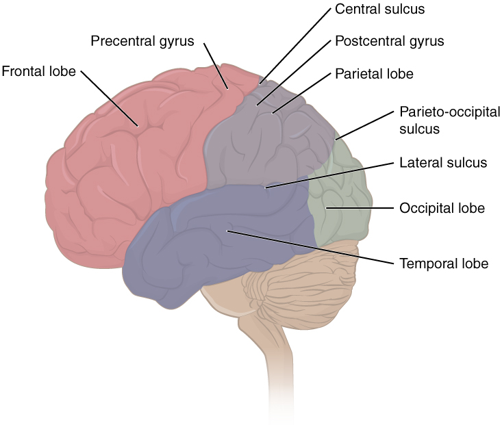
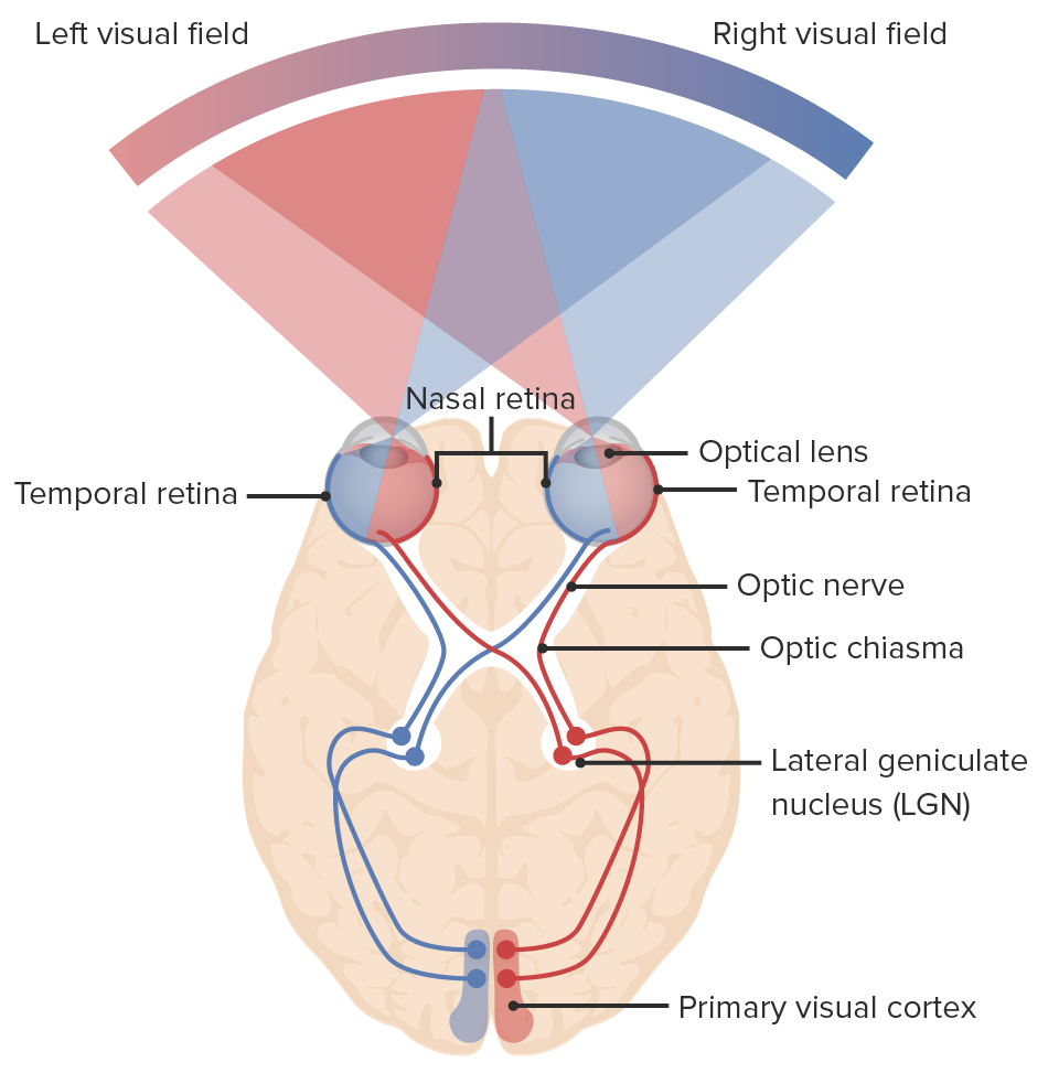
00:00 Now, we have come upon the visual pathway itself. The primary visual cortex is in the occipital lobe of the cerebrum. So to visualize, to perceive vision, light has to go through a fairly complicated visual pathway to get to this primary visual cortex. Let’s take that particular journey. 00:31 The first thing to kind of consider here is that we have two visual fields. We have a left visual field over here. We have a right visual field over here. Another consideration is the arrangement of the retinae in this pathway. We are going to define the retinae as having a temporal component as well as a retinal component. So if we look in the left eyeball of the left retina, this more medial aspect is close to the nose. So this is the nasal retina of the left eye and then this will be the temporal retina of the left eye. Over here, we have the nasal retina of the right eye. 01:31 We have the temporal retina of the right eye. The temporal retina, when the photoreceptors there are activated, optic nerve fibers are going to travel ipsilaterally into the optic tract. 01:50 So let’s take a look at how those fibers are travelling. Temporal retina here in the left eye and then when this retina is activated, we see the optic nerve. We see the optic chiasm. 02:08 Then these fibers from the temporal retina will stay ipsilaterally to then travel to the left primary visual cortex. Same thing with the right eye except the temporal retina here is in red. 02:24 The red fibers travel here in the optic nerve, get into the optic chiasm, do not cross and then continue their journey ipsilaterally to the primary visual cortex. This is in contrast to the path that nerve fibers take from the nasal retina. If we take a look here, here’s the nasal retina associated with the left eye. If we follow the nerve fibers from the nasal retina of the left eye, they come into the optic nerve. They get to the optic chiasma. Instead of staying on the same side, they crossover at the optic chiasma, and thus become contralateral and then end up in the contralateral primary visual cortex, which should be the right one. If we take a look at the right nasal retina over here and follow its nerve fibers to enter the optic nerve, come to the optic chiasma and then crossover to the left side. Then they terminate ultimately in the left primary visual cortex. The temporal retina is on the lateral side, those fibers run ipsilaterally. 03:46 The nasal retina on the medial aspect, those fibers cross within the optic chiasma. 03:53 Now, we need to continue our journey. The fibers will synapse within the lateral geniculate body that we see here. Over here, that would be most of the fibers. Some of the fibers coming from the retinae will project to other neural structures including the superior colliculi, the pretectal area as well as the suprachiasmatic nucleus. But we’re not going to focus on those projections. We’re going to continue our journey then from the lateral geniculate body to the primary visual cortex. What will happen here is neurons from the lateral geniculate body will travel in optic radiations. These fibers here running from your lateral geniculate body going to the primary visual cortex are your optic radiations. They are identified over here on the right as well. That is the journey that is taken in order for us to perceive vision.
About the Lecture
The lecture Primary Visual Cortex by Craig Canby, PhD is from the course Visual Pathways.
Included Quiz Questions
How do optic nerve fibers from the temporal and nasal retina travel into the optic tract?
- Fibers from the temporal retina travel ipsilaterally into the optic tract, whereas those from the nasal retina travel contralaterally.
- Fibers from both the temporal and nasal retina travel ipsilaterally into the optic tract.
- Fibers from the right temporal retina travel ipsilaterally into the optic tract, whereas those from the left temporal retina travel contralaterally.
- Fibers from the temporal retina travel contralaterally into the optic tract, whereas those from the nasal retina travel ipsilaterally.
- Fibers from both the temporal and nasal retina travel contralaterally into the optic tract.
At which of the following structures do most of the optic nerve fibers coming from the retina synapse before entering the primary visual cortex?
- Lateral geniculate nucleus
- Pretectal area
- Superior colliculi
- Edinger-Westphal nucleus
- Suprachiasmatic nucleus
Author of lecture Primary Visual Cortex

Craig Canby, PhD
Customer reviews
5,0 of 5 stars
| 5 Stars |
|
5 |
| 4 Stars |
|
0 |
| 3 Stars |
|
0 |
| 2 Stars |
|
0 |
| 1 Star |
|
0 |
