Playlist
Show Playlist
Hide Playlist
Prenatal Screening
-
Slides 03 Genetic Counseling and Prenatal Diagnosis PopulationGenetics.pdf
-
Download Lecture Overview
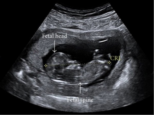
00:01 Stepping back a little bit, some of the techniques that you may be involved in as a doctor before referring to a genetic counselor are prenatal screening diagnosis techniques. 00:14 Again, most of these are not actually genetic tests. They’re tests that might come before we would refer for any particular genetic testing. Prenatal screening is non-invasive and used to identify the need for perhaps more invasive prenatal diagnosis techniques. 00:34 But I have to say there are some new technologies even so on the forefront that really open our eyes to perhaps not even having to use some of the more invasive techniques. 00:46 In the battery of things that we can do in prenatal screening, we can simply take blood measurements of analytes, so proteins and hormones and things that we might find in the maternal serum. 01:00 Probably, you’re already familiar with alpha-fetoprotein. MS-alpha-fetoprotein is alpha-fetoprotein from the maternal serum. We can measure this as you see in this figure. Take a moment to think what that figure is showing you. You can see the X axis is saying what the maternal serum for alpha-fetoprotein levels are and also the proportion of individuals that are unaffected. 01:33 On the very left side, you have Down syndrome or on the right side, fetuses that have spina bifida. 01:40 You can see that measuring maternal serum levels of alpha-fetoprotein, if an individual fetus is showing or maternal serum is showing high levels of alpha-fetoprotein, it could indicate spina bifida or low levels could indicate Down syndrome. Now, these are not exclusive tests. As you see, there’s quite an overlap between the unaffected individuals and Down syndrome individuals. 02:10 We need to use additional tests like ultrasound screening in order to verify that maybe there’s a neural tube defect in the event that we see high levels of maternal serum alpha-fetoprotein or to see some of the other signs, phenotypic signs of Down syndrome like smaller hands and such. 02:31 Another measurement that we can measure is hCG. Do you remember what hCG is? Probably you do, human chorionic gonadotropin. Not only does hCG increase and show whether an individual is pregnant, whether a mother is pregnant, one of the first cues we measure is for human chorionic gonadotropin, right? But if it’s particularly elevated, it could also indicate trisomy 21 or Down syndrome. We could use that in conjunction with the alpha-fetoprotein measurements in maternal serum. This though is the exciting one that I wanted to tell you about, cell free DNA. I’m not sure how many of you have heard of cell free fetal DNA testing before because it’s a relatively new technique and certainly exciting because it allows us a non-invasive prenatal screening mechanisms simply by testing the maternal blood. Measuring fetal cell free DNA in the maternal blood is a really cool technique and relatively new. We call it NIPS for non-invasive prenatal screening. Through the analysis of fetal cell free DNA in the maternal blood, we can actually make a very accurate prediction of aneuploidies. Let me show you how this works because it’s a little bit complex. But I’m pretty sure even if you haven’t heard of it before, I can lead you into a great understanding. Let’s say to start with that there are millions of particles of DNA. 04:25 These particles of fetal DNA start appearing as early as about seven weeks of pregnancy. 04:32 So, we can get not only prenatal diagnosis but early prenatal diagnosis for some of these disorders. 04:39 These millions of particles of DNA that will be extracted in the maternal blood will be mapped to their chromosome of origin because they get tagged and what not. So, we’ll have many, many, many sequences of different pieces that based on their appearances belong on certain chromosomes. 05:01 Then these pieces can be matched up. Basically, we will get a read out of how many pieces of chromosome 1, 2, 4, whichever there are. This is just a subset of the representation here. 05:23 Regardless of whether they’re fetal or maternal origin, if the fetus happens to have an additional chromosome, we will see proportionally higher representation of the DNA associated with that chromosome. Then we take these sequences that have been aligned to their chromosome number. Of course, computer applications really help with this. 05:49 Then we can see a graph representing how much DNA there is for each chromosome. 05:59 This is compared to standards of what we would expect for the contribution to each of the chromosomes. 06:06 If there is extra DNA showing up then we can make a prediction that there are perhaps extra copies or extra fragments of chromosome. This is particularly useful in diagnosing the aneuploidies like Down syndrome, Patau syndrome, Edwards syndrome as well as the sex chromosome aneuploidies that we might run into. I mentioned in addition to some of these nongenetic tests, the cell free DNA of course was a genetic test because we’re looking at the DNA. But the previous ones were not genetic tests and nor is ultrasonography. I’m sure you are very familiar with ultrasound also. 06:49 It can be used in addition to these blood sampling techniques in order to confirm or release us from suspicion of any genetic abnormality. For example, we might test for neural tube defects. 07:06 You can see here in the left, there’s a normal skin covering. Then on the right side image, there’s a bulge in which we can see that the neural tube has not sealed. Now of course, you are not going to be an expert nor am I at reading ultrasonography or ultrasonograms. 07:28 That’s not the point here. The point is that the technique can be used in order to confirm a genetic disorder.
About the Lecture
The lecture Prenatal Screening by Georgina Cornwall, PhD is from the course Population Genetics.
Included Quiz Questions
Which of the following is the most sensitive noninvasive prenatal screening test for Down syndrome?
- Cell-free DNA testing
- Fetal villus sampling
- Targeted ultrasound
- Amniocentesis
- Quadruple test
Which of the following changes in maternal serum markers may indicate Down syndrome in the fetus?
- Reduced alpha-fetoprotein, elevated human chorionic gonadotropin
- Elevated alpha-fetoprotein, reduced human chorionic gonadotropin
- Reduced alpha-fetoprotein and human chorionic gonadotropin
- Elevated alpha-fetoprotein and human chorionic gonadotropin
- Maternal serum markers for Down syndrome in the fetus do not include alpha-fetoprotein or human chorionic gonadotropin.
High levels of maternal serum alpha-fetoprotein may indicate which of the following conditions in the fetus?
- Spina bifida
- Down syndrome
- Alpert syndrome
- Treacher Collins syndrome
- Trisomy 18
Which diagnostic tool is most commonly utilized to visualize fetal neural tube defects?
- Ultrasound
- Radiography
- Magnetic resonance imaging
- Computed tomography scan
- Fetal villus sampling
Customer reviews
5,0 of 5 stars
| 5 Stars |
|
5 |
| 4 Stars |
|
0 |
| 3 Stars |
|
0 |
| 2 Stars |
|
0 |
| 1 Star |
|
0 |


