Playlist
Show Playlist
Hide Playlist
Posterior Compartment – Anatomy of the Leg
-
Slides 06 Lower Limb Anatomy.pdf
-
Download Lecture Overview
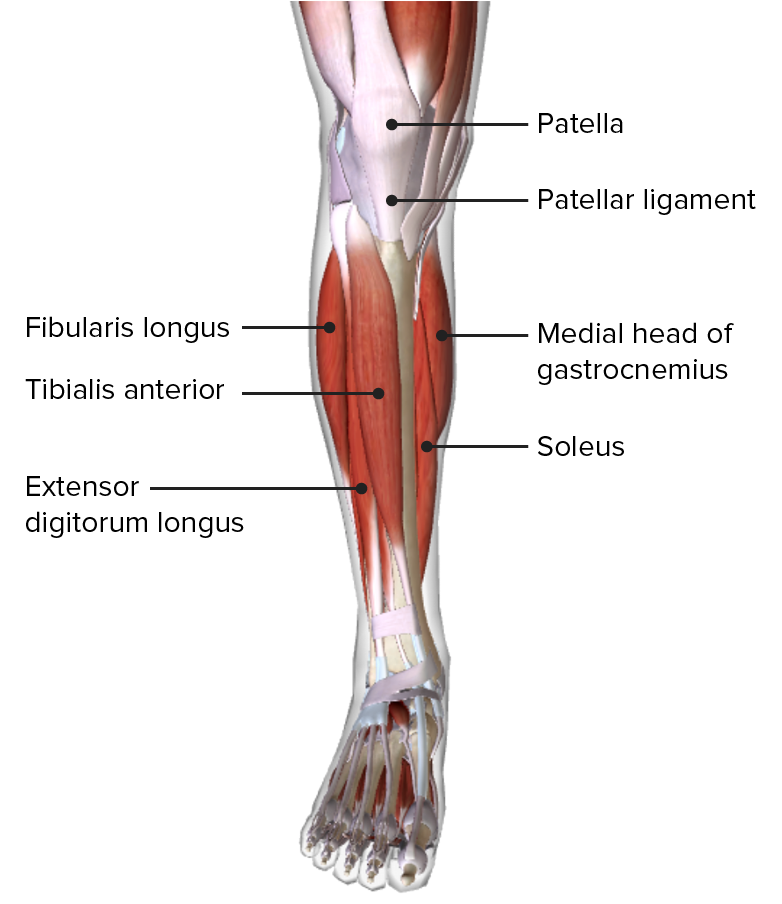
00:00 If we now move on to the posterior compartment of the leg, we can see we have both superficial and deep layers that I mentioned. Let’s deal with the superficial layers first. 00:11 On here, we can see most superficially closest to the skin, we have gastrocnemius. And this has two heads. There’s a lateral head to gastrocnemius we can see running over here. 00:26 This is a lateral head of gastrocnemius, and here we can see the medial head of gastrocnemius. 00:33 So here, we have the posterior view of a right leg. We have the lateral head of gastrocnemius here, and we have the medial head here. Deep to gastrocnemius, we find we have soleus, and this is a large muscle, a very fleshy muscle that is sitting directly deep to gastrocnemius. 00:53 So gastrocnemius, the lateral head, comes from the lateral condyle of the femur while the medial head comes from the medial condyle of the femur. Soleus, this comes from the posterior surface of the fibula, and also as we mentioned in the osteology lecture, it comes from the soleal line of the tibia. Both of these muscles pass through the posterior aspect of the calcaneus via the calcaneal tendon. They’re supplied by the tibial nerve. 01:24 So these muscles in the posterior compartment are supplied by the tibial nerve, one of those divisions that are coming away from the sciatic nerve. Gastrocnemius and soleus are both involved in plantarflexion of the ankle, so enabling you to stand on tiptoes. And because gastrocnemius crosses the knee joints, then it can actually flex the knee as well. One other muscle that I haven’t mentioned is plantaris. Plantaris is running alongside the medial aspect of the lateral head, and this gives rise to a very thin and long tendon that runs between gastrocnemius and soleus. So we can see plantaris. It’s coming from the inferior aspect of the lateral supracondylar ridge of the femur, and its long tendon passes between the soleus muscle and the gastrocnemius muscle. It has a long tendon that then blends with the calcaneal tendon inserting on to the calcaneus. It’s in the posterior compartment. So it’s also supplied by the tibial nerve, and it is a weak plantarflexor of the ankle. If we look at more deep layers, then there are three muscles here that I want to talk about, first of all. We have popliteus, flexor digitorum longus, and flexor hallucis longus. 02:48 So we can see these if we look into the deep compartments. We have popliteus here. 02:54 We have flexor digitorum longus. We have flexor hallucis longus. 02:59 So popliteus, we can see coming from these lateral aspects of the leg. It’s coming from the lateral surface of the femoral condyle. 03:08 We can see it’s passing across to the tibia. We then have long muscles that give rise to tendons that pass into the foot, flexor digitorum longus and flexor hallucis longus. So we can see popliteus coming from the lateral aspects of the lateral condyle of the femur. It also comes from the lateral meniscus, and it passes to the posterior tibia above the soleal line which we mentioned before. Flexor digitorum longus, this is coming from the posterior surface of the tibia below the soleal line, and this passes to the distal phalanges of digits 2 to 5. Flexor hallucis longus, this is coming from the lower two-thirds of the posterior fibula, and also their interosseous membrane. It passes to the distal phalanx of the great toe. All of these muscles, as they’re in the posterior compartment, are supplied by the tibial nerve. Popliteus, this is going to be a weak knee flexor. 04:11 It’s also involved in unlocking the knee by lateral rotation of the femur on a stable tibia. 04:19 So when you’re in standing position and the femoral condyle is tightly articulating with the tibial plateau, then popliteus is important in unlocking the knee enabling flexion to occur. 04:36 Flexor digitorum longus flexes the digits 2 to 5, and it also helps to plantarflex the ankle. Flexor hallucis longus flexes the great toe and is also a weak plantarflexor. 04:49 There’s a fourth muscle in the deep layer of the posterior compartment of the leg. 04:55 That is the Tibialis posterior. It originates as 2 bands on the posterior surface of the tibia, fibula, and interosseous membrane and then merges into 1 muscle mass half-way down the leg. 05:05 At the ankle, the tibialis posterior passes between the medial malleolus of the tibia and continues on the medial side of the foot, passing under the flexor retinaculum. 05:16 In the foot, the tibialis posterior inserts into the tuberosity of the navicular bone, medial cuneiform bone, and the bases of the second, third, and fourth metatarsal bones. 05:27 This muscle is the main invertor of the foot and also aids in plantarflexion as well as supporting the plantar arches.
About the Lecture
The lecture Posterior Compartment – Anatomy of the Leg by James Pickering, PhD is from the course Lower Limb Anatomy [Archive].
Included Quiz Questions
Which muscle forms the bulk of the muscle in the posterior lower leg?
- Gastrocnemius
- Flexor hallucis brevis
- Flexor hallucis longus
- Flexor digitorum brevis
- Soleus
What is the site of origin of the soleus muscle?
- Posterior surface of the fibula
- Anterior supracondylar ridge of the femur
- Lateral supracondylar ridge of the femur
- Posterior supracondylar ridge of the femur
- Anterior surface of the fibula
What is the most superficial muscle in the posterior compartment of the leg?
- Gastrocnemius
- Popliteus
- Soleus
- Fibularis longus
- Tibialis anterior
Which muscle of the posterior compartment of the leg inserts on the distal phalanx of the hallux?
- Flexor hallucis longus
- Plantaris
- Popliteus
- Gastrocnemius
- Soleus
Which muscle is responsible for inversion of the foot?
- Tibialis posterior
- Plantaris
- Popliteus
- Gastrocnemius
- Soleus
Customer reviews
5,0 of 5 stars
| 5 Stars |
|
5 |
| 4 Stars |
|
0 |
| 3 Stars |
|
0 |
| 2 Stars |
|
0 |
| 1 Star |
|
0 |


