Playlist
Show Playlist
Hide Playlist
Physical Findings in Knee Pain

-
Reference List Physical Examination.pdf
-
Download Lecture Overview
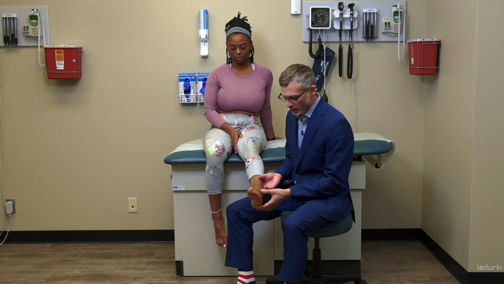
00:01 Having assessed the menisci, we can now move on to assess two other conditions that are relatively common. 00:08 I'm going to first look at patellofemoral pain syndrome. 00:10 This is particularly common in young women. 00:14 And essentially it's an overzealous Q angle where they're even more valgus than the baseline. 00:21 And that's because of some imbalance in the different ligamentous structures holding the knee together. 00:26 And oftentimes it's a problem with too much activation of the quadriceps, the vastus lateralis And so if the knee is a little bit too inward, and so what I'm looking for is to see if there's any pain associated with the kneecap itself and how it's moving in the patellofemoral groove. 00:45 So what we're going to do is put your knee at around 30 degrees and I'm simply trying to bring your kneecap towards me. 00:52 Patients who have patellofemoral pain syndrome will have a little bit of instability around the patella. 00:58 And by bringing it towards me, the patient will have what's called apprehension, an apprehension sign where she'll either put her hand up and say, "stop, that's uncomfortable, I feel like you're going to knock my knee off or just or dislocate my knee" or they'll just immediately bring the leg down like this as sort of a reflex instinct that would prevent me from dislocating her knee. 01:19 And that's because if I flip over my kneecap here, your patient is having discomfort to the lateral aspect of the knee because it's chronically displaced laterally. 01:28 We'll have a lot of discomfort when I bring that kneecap laterally in that position. 01:33 So that's called the patellofemoral apprehension sign. 01:37 Next up, we'll do a test for the iliotibial band. 01:41 I'll have you roll over onto your left hip again, please. 01:47 So the iliotibial band starts up here with the tensor fascia lata, and it's coming down here and inserting onto Gerdy's tubercle down here by the fibula. 01:57 And if you could just lift up your leg, if I pushed down and have her resist me, I'm going to be able to feel very easily this tight band here called the iliotibial band. 02:06 It's a very firm structure of musculotendinous tissue, and it's inserting down here. 02:13 You can relax now. 02:14 And if I want to try and see if the iliotibial band is inflamed, I can put my thumb. 02:20 I'm going to show it on my model here again. 02:22 I put my thumb right here just proximal to the lateral epicondyle. 02:27 In this position, the iliotibial band is anterior to my thumb, but as soon as I flex her knee, the iliotibial band ends up behind my thumb. 02:39 So keeping my thumb here, by moving her knee between flexion and extension, the iliotibial band will pass underneath my thumb repeatedly. 02:48 And if the patient has pain associated with iliotibial pain syndrome, this will reproduce that pain as the iliotibial band passes to and fro underneath my thumb. 02:58 And this is called 'Noble's test'. 03:01 Noble's test is at the knee. 03:03 So that's one way that I remember that test. 03:07 The last test we're going to do, I'll have you go again on to your back one last time, is even in the setting of minor trauma, patients sometimes can have a tibial plateau fracture or a tibial insufficiency fracture and simply percussing along the tibial like so to see if any specific point causes any discomfort may be an indication of a tibial insufficiency fracture.
About the Lecture
The lecture Physical Findings in Knee Pain by Stephen Holt, MD, MS is from the course Examination of the Lower Extremities.
Included Quiz Questions
What does the patellar apprehension test evaluate for?
- Patellofemoral instability
- Pes anserine bursitis
- Iliotibial band syndrome
- Tibial insufficiency fracture
- Knee effusion
What might exquisite tenderness on palpation of the anterior tibia indicate?
- Tibial insufficiency fracture
- Iliotibial band syndrome
- Patellofemoral instability
- Pes anserine bursitis
- Anterior cruciate ligament tear
Author of lecture Physical Findings in Knee Pain

Stephen Holt, MD, MS
Customer reviews
5,0 of 5 stars
| 5 Stars |
|
1 |
| 4 Stars |
|
0 |
| 3 Stars |
|
0 |
| 2 Stars |
|
0 |
| 1 Star |
|
0 |
Great course detailed and profound explanation easy to memorize and use in daily practice.Thank You.
