Playlist
Show Playlist
Hide Playlist
Physical Exam Findings in Spontaneous Bacterial Endocarditis

-
Reference List Physical Examination.pdf
-
Download Lecture Overview
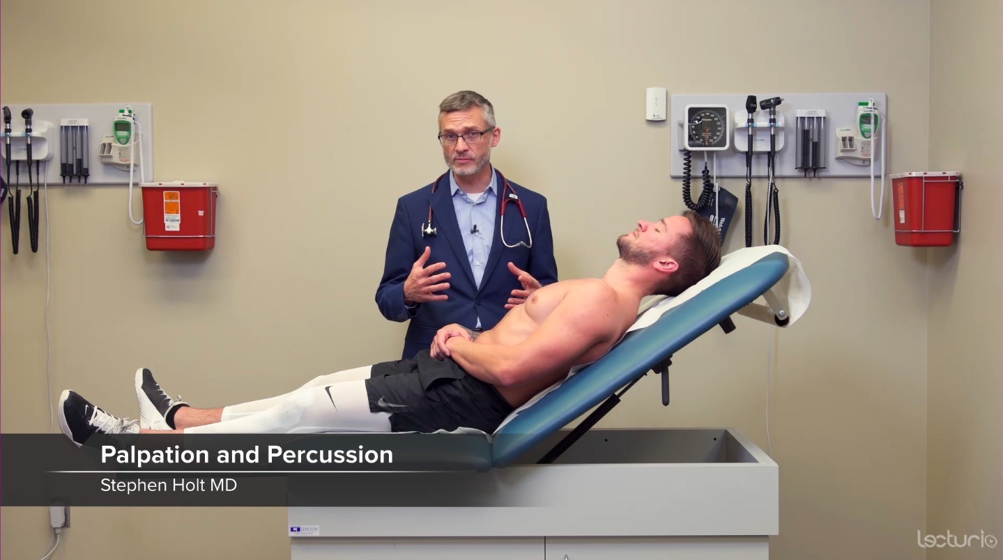
00:01 So the last condition that I wanted to touch upon in terms of the cardiovascular system, is the assessment of endocarditis, most specifically, spontaneous bacterial endocarditis. 00:10 In addition to listening for a murmur which I was talking before about if you have acute or new tricuspid regurge or a new murmur anywhere amongst the heart valves, you should be thinking about endocarditis particularly on the patient with the risk factors and the fever and positive blood cultures. 00:26 But there's a few common stigmata of spontaneous bacterial endocarditis that are worth being aware of. 00:33 These are all essentially manifestations of micro seeding or little micro emboli of septic material from the heart valve shooting off into other parts of the body. 00:44 When you have right-sided heart disease like the tricuspid valve, those little seeds are only going to go towards the lungs and may cause little septic emboli, little tiny pneumonias, so to speak, in the chest wall. 00:56 However, in patients with right-sided heart disease, those little micro emboli of septic material are shooting off into the entire population of your blood stream and therefore they can show up in end arteries. 01:10 Now in particular, the ones we were looking -- we'd for in the hands would be splinter hemorrhages which are evidence of little microthrombi that have broken off that are just made manifest in the nail bed. 01:22 On the other side of the hand, we might see Janeway lesions which are painless lesions that oftentimes appear on the palms or on the hands, they are violaceous, potentially necrotic appearing in color, and again they are painless. 01:39 And then there is Osler's nodes, and Osler's nodes may be on the fingers, again, anything that's happening on the fingers can also occur in the toes or on the soles. 01:47 Osler's nodes in contrast to Janeway lesions are painful. 01:51 They also are evidence of either vasculitis or sometimes they are able to culture bacteria from them so there's some evidence that they are micro emboli of bacterial colonies and you can see them on joints over the hands, occasionally on the palms and soles as well. 02:07 The last place you might look for these stigmata of endocarditis would be rough spots in the back of the eye, but we're not going to talk about the retina anymore today.
About the Lecture
The lecture Physical Exam Findings in Spontaneous Bacterial Endocarditis by Stephen Holt, MD, MS is from the course Examination of Cardiovascular and Respiratory System.
Included Quiz Questions
What are splinter hemorrhages seen with bacterial endocarditis?
- Microthrombi in the nail bed
- They are also known as Janeway lesions.
- They are also known as Osler nodes.
- Circumscribed painful erythematous swelling in the skin and subcutaneous tissues of the hands
- Small hemorrhages seen in the retina
Author of lecture Physical Exam Findings in Spontaneous Bacterial Endocarditis

Stephen Holt, MD, MS
Customer reviews
5,0 of 5 stars
| 5 Stars |
|
5 |
| 4 Stars |
|
0 |
| 3 Stars |
|
0 |
| 2 Stars |
|
0 |
| 1 Star |
|
0 |
