Playlist
Show Playlist
Hide Playlist
Photoreceptors: Rods and Cones – Vision (PSY, BIO)


-
Slides Vision SensingtheEnvironment.pdf
-
Download Lecture Overview
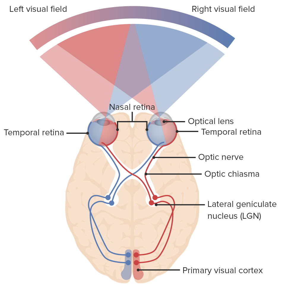
00:01 So take a look at this image. 00:03 On your left, we have a nice sunny day and you can see color and light and everything seems really great. 00:08 And then we know that dusk time at night where things start to get dark. 00:11 On the right you notice, you don’t see color. 00:14 All you see is borders and shade, but you don’t see clear color, right? And that is because of the differences between what’s being activated in terms of the rod cells versus cone cells. 00:24 So color is cones and darkness and light detection is rods. 00:30 So, here is an image illustrating the difference between the two. 00:35 We can see where it gets its name. 00:36 The rod has this rod shaped, you know, cricket or baseball bat formation. 00:41 And the cone, I’m thinking ice cream cone upside-down. 00:45 So they have this unique shape, which is where they get their name. 00:48 And you look at where the lights, so the arrow at the top of the diagram is showing where the light interacts with these cells. 00:54 And there are certain features that they share. 00:56 So they both have a nucleus, they both have mitochondria which is an energy source. 01:00 And they have an outer segment that contains this chemical and this photosensitive chemical or pigment is what allows us to actually engage and detect this light. 01:11 So let’s get in to more detail around that. 01:14 First, we’ll take a look at sort of the characteristics of rods versus cone. 01:17 So rods or scotopic vision is where we have these rod cells being activated. 01:23 Now in the human retina, we have about 120 million rod cells. 01:29 And for sure, the MCAT exam is going to make you, force you, one way or the other, to differentiate between which one is more abundant. 01:36 And that would be the rods and maybe even numbers. 01:39 So 120 and we’ll see how many cones are in just a sec. 01:42 So they’re strongly photosensitive, meaning light dark. 01:45 So less detail, no color, concentrated at the periphery. 01:49 So they’re not at the fovea centralis, they’re more around the edges of the eye, okay? So they’re using low light conditions. 01:55 So a little, you know, a little, I don’t want to say trick, but a little, a little behavior you can do is when you’re out and it is dark, if you’re looking directly at something that’s dark and that’s what you’re trying to detect, you won’t see it very well. 02:08 But if you move your eye a little bit to the side, you’ll actually be bringing the light to the edges where a lot of these rod cells are actually focused. 02:17 A lot of astronomers when they’re looking at stars and they’re having trouble detecting a star, look a little bit askew to the star that you’re actually trying to detect and you’ll see it a little bit better. 02:28 Cone cells are found a lot less abundantly. 02:31 It’s around six million. 02:33 And they’re weakly photosensitive so they are okay with light, not great. 02:37 They’re more about like high acuity, more resolution, more detail and specifically color. 02:42 So in order to remember cones, color that sees, I think that might help you. 02:46 Concentrated at the fovea like we’ve mentioned and they’re great at bright conditions, okay? So, both respond to light and there has to be something else that they have in common that allows them to sort of do their function in a similar fashion. 03:03 Because really, I mean, they sort of look the same but they have all these different properties. 03:06 They’re both responding to light, but what’s driving that difference? Well, the difference lies into the actual processing and some of the proteins that they contain. 03:14 So they each have a pigment protein called opsin that changes in response to light. 03:19 So it’s a photosensitive pigment. 03:22 Now opsin contains a pigment molecule called retinal, which is expressed as one of two things depending on which cell you’re in. 03:29 The rods have rhodopsin. 03:31 So that might help you remember. 03:32 Row, rhodopsin, rod, right? So rhodopsin and rod cells and photopsin in cone cells. 03:39 So depending on the presence of which molecule is there, that will determine what type of processing is going to happen. 03:45 So at rest, opsin keeps the sodium channels open making the cell depolarize which causes glutamate release. 03:52 Okay? So we should kind of try and grasp that. 03:54 At rest, opsin keeps the sodium channels open keeping the cell depolarized. 03:59 And if you remember from our previous lectures that we’ve done, we were talking about resting membrane potential and depolarization versus hyperpolarization. 04:08 Now phototransduction is the process of how we convert this photon of light into actual information. 04:14 So the photon of light converts retinal from its all-trans form. 04:18 So again, we don’t want to go deep into chemistry but you can have a “trans” or “cis” form of a molecule. 04:24 So everything right now would be all transformed and closes the sodium channels, which hyperpolarizes the cell, okay? So light, converts retinal to all-trans form and hyperpolarizes the cell. 04:35 Glutamate then inhibits bipolar cells. 04:37 And once the hyperpolarization is removed by the inhibition, this activates the ganglion cells, okay? So activation of the ganglion cells then together sends information through the optic nerve to the brain. 04:51 Now the optic nerve does a lot of different really cool things. 04:53 It can activate and -- a lot of different parts of your brain. 04:58 They do different things other than just straight visual acquisition. 05:02 But for our discussion here, we’re talking about how this optic nerve projects to the visual centers of the brain.
About the Lecture
The lecture Photoreceptors: Rods and Cones – Vision (PSY, BIO) by Tarry Ahuja, PhD is from the course Sensing the Environment.
Included Quiz Questions
Compared to the cones, the rods are...? (Select all that apply)
- ...much more numerous.
- ...capable of showing color in bright light.
- ...less sensitive to light.
- ...capable of revealing more detail in an image.
- ...concentrated at the periphery of the retina.
What is the best method to look at stars (as pertaining to photoreceptors)?
- With the head directed slightly off-center
- With the head directed straightforward
- With the head directed straightforward through a pinhole
- With the head directed completely to the side and extended completely
- With the head directed completely to the side
Compared to the rods, the cones have...? (Select all that apply)
- ...a shorter length.
- ...a different signaling molecule than cGMP for phototransduction.
- ...color-sensitive pigments.
- ...a different response to light of different wavelengths.
- ...the most numbers in the fovea, towards the central portion of the retina.
In which situation would excitation of rods occur most often?
- While trying to find a light switch in the dark
- While on a picnic
- While in a store
- While hiking
- While driving
Author of lecture Photoreceptors: Rods and Cones – Vision (PSY, BIO)

Tarry Ahuja, PhD
Customer reviews
5,0 of 5 stars
| 5 Stars |
|
5 |
| 4 Stars |
|
0 |
| 3 Stars |
|
0 |
| 2 Stars |
|
0 |
| 1 Star |
|
0 |
