Playlist
Show Playlist
Hide Playlist
Patient Introduction and Review of Breast Anatomy and the Lymph System

-
Slides Physical Exam Breast Lump-Introduction.pdf
-
Reference List Physical Examination.pdf
-
Download Lecture Overview
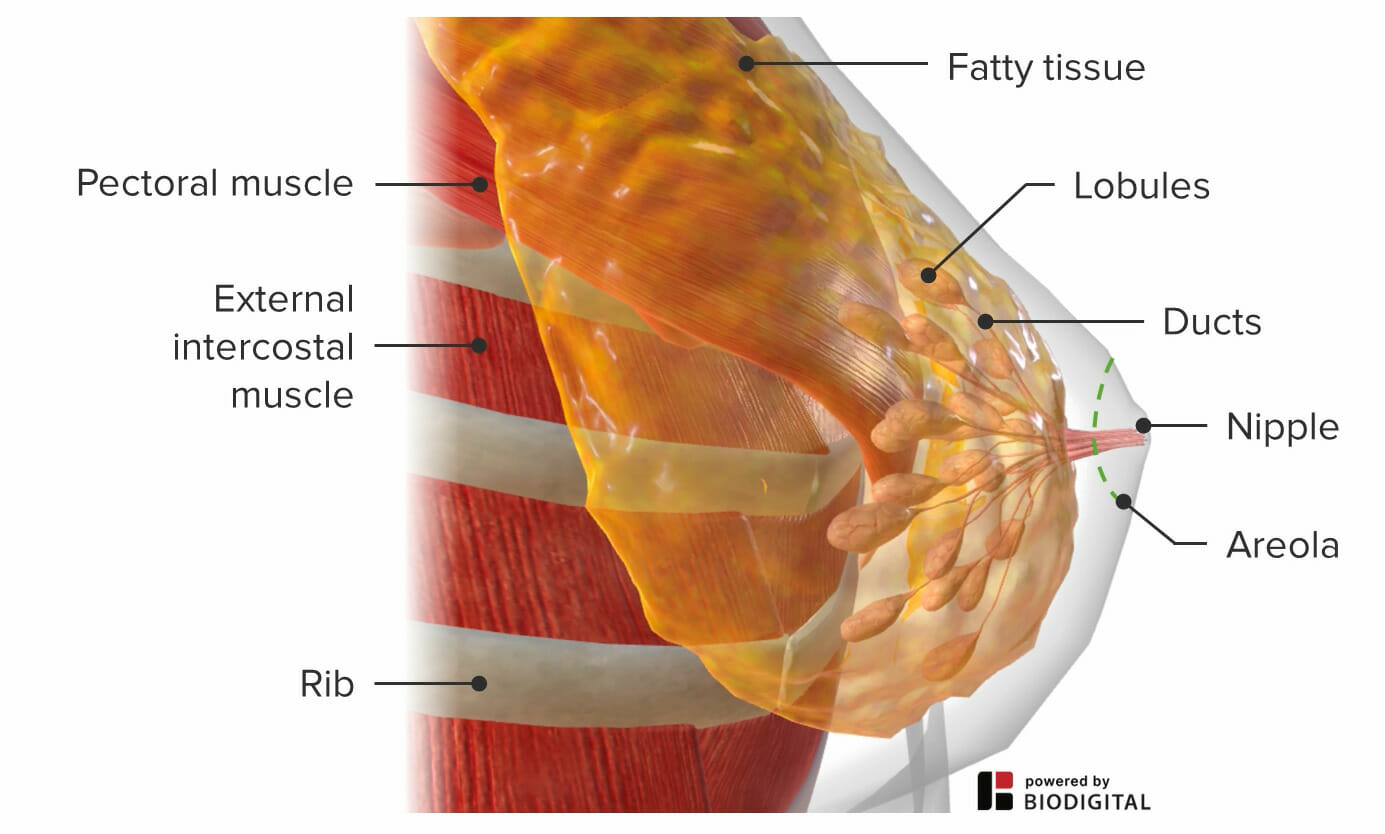
00:01 So let's now move on to the evaluation of a breast lump. 00:05 This is a 44-year-old woman who's presenting with a lump in her right breast. 00:09 She first noticed it about 4 months ago and says that it is painful just before menstruation. 00:15 So we're going to talk about the difference between benign versus malignant breast lumps, and we'll go through some basic features of these two different kinds of lumps. 00:24 And then, of course, we'll move on to the physical exam, which will be important in our assessment. 00:29 So typically benign breast lumps are slow growing. 00:33 They tend to be painful or they're more likely to be painful. 00:37 They're encapsulated, which means that they typically have a nice, smooth contour with symmetry and they're rubbery. 00:45 You can push on them and they will compress. 00:47 They're not rock hard. 00:49 In contrast, malignant tumors tend to be faster growing. 00:52 They are most often painless unless very advanced. 00:56 They're not encapsulated so they don't have smooth edges. 01:00 They're much firmer, of course. 01:02 And importantly, we'd be looking for signs of lymphadenopathy if there's evidence of metastatic spread. 01:10 Quick review of the anatomy of the breast, remember that most of the breast is composed of fatty tissue. 01:15 And then heading towards the nipple, we have lobules that are emptying into ducts and then the ducts converge at the areola and exit via the nipple. 01:27 Common benign breast tumors include fibroadenomas, which are most common between the ages of 15 and 35. 01:34 These do not change with menstruation and they can be brought on by oral contraceptive pills. 01:41 In contrast, fibrocystic changes are very common. 01:44 They tend to be exacerbated around the time of menses. 01:48 So women will often report that they have more painful breasts prior to menses and then they can resolve after the menstrual period is over. 01:56 And thirdly, Galactoceles. 01:58 These occur in women of childbearing age. 02:00 They are relatively firm subareaolar masses. 02:04 In contrast, here we have the malignant breast cancers. 02:08 Ductal carcinoma, which are the most invasive types of breast cancer. 02:12 Lobular carcinoma, which oftentimes presents in both breasts simultaneously. 02:17 Paget's disease. 02:19 And then inflammatory carcinoma, which can actually look all the world like cellulitis or mastitis involving the breast. 02:27 Which is a good transition to talk about infectious manifestations for breast enlargement. 02:32 Mastitis, particularly in women who are breastfeeding, and then the progression of mastitis can lead to an actual abscess with fluctuants within the wall of the breast. 02:44 In the assessment of any patient with a breast lump, it's going to be very important to make sure we do a thorough lymphatic exam. 02:50 This will be useful for screening, for malignancy and of course, also for infection. 02:55 In general, lymph nodes are expected to be less than a centimeter in size to be normal. 03:00 However, in the axilla, we'll actually tolerate up to 2 or 3 centimeters in size before we would get too concerned. 03:07 You'll always want to be comparing one side to the other as symmetry is important when we're comparing the different sizes of lymph nodes that we can palpate. 03:16 Honing in on the lymphatic exam of the head and neck, there are predictable regions where lymph nodes tend to come together and we may or may not be able to palpate them in asymptomatic people, but it's important to know where to look for them. 03:29 And we'll be covering those details more when we move on to the physical exam. 03:34 Moving down from the head and neck, we'll remind ourselves that the lymphatic systems follow predictable lines of drainage, with both extremities emptying into the great vessels of the neck, but the lower extremities draining into the thoracic duct, which then will drain into the left great vessels. 03:52 We were just talking about the size of lymph nodes. 03:54 And as I mentioned, in general, a lymph node is more concerning if it's greater than 2 centimeters in size, if it's growing very quickly over time, if there's asymmetry, the mobility of the lymph node can also correlate with the extent of concern that we should have. 04:09 Those that are mobile are less likely to be malignant. 04:13 And lastly, lymph nodes that are tender are more likely to be associated with infection than malignancy. 04:21 So returning to our case of our middle aged woman with breast pain, things that are on our differential would include fibrocystic changes, a fibroadenoma, breast cancer, of course, galactocele and mastitis.
About the Lecture
The lecture Patient Introduction and Review of Breast Anatomy and the Lymph System by Stephen Holt, MD, MS is from the course Examination of the Breast and Lymph System.
Included Quiz Questions
A breast mass in a 25-year-old woman described as small, firm, mobile, and unchanging with menstruation is likely what type of pathology?
- A fibroadenoma
- Breast cancer
- Fibrocystic change
- A galactocele
- Paget's disease
Which characteristic of an axillary lymph node favors a benign process?
- Size < 2 cm
- Non-tender
- Rapidly growing
- Asymmetric compared to the contralateral axilla
- Non-mobile
Author of lecture Patient Introduction and Review of Breast Anatomy and the Lymph System

Stephen Holt, MD, MS
Customer reviews
5,0 of 5 stars
| 5 Stars |
|
5 |
| 4 Stars |
|
0 |
| 3 Stars |
|
0 |
| 2 Stars |
|
0 |
| 1 Star |
|
0 |
