Playlist
Show Playlist
Hide Playlist
Paget's Disease with Case
-
Slides Calcium Metabolis.pdf
-
Reference List Endocrinology.pdf
-
Reference List Metabolic Bone Disorders.pdf
-
Download Lecture Overview
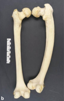
00:01 In this case, a 67-year old man with prostate cancer since the age of 63 comes to see you. 00:07 His only symptom is new-onset upper back pain. 00:11 He takes no medication. 00:12 On physical examination, his vital signs are normal. 00:16 His spine is tender to palpation over the thoracic area. 00:19 Lab studies show serum calcium level of 9.6 mg/dL, a serum creatinine level of 1.0 mg/dL A serum Alk Phos of 240 and a PSA of less than 4 ng/mL. 00:34 Whole body radionucleide bone scan shows focal increase uptake at T7. 00:40 There are no other abnormalities. 00:43 The spine x-ray shows coarsening of trabeculae and expansion of the body of T7 without cortical disruption. 00:52 What is the most likely diagnosis? In this scenario, we have an elderly patient with a history of prostate cancer presenting with back pain and point tenderness over the spine. 01:03 In ruling out bone metastases, he has an incidental finding of coarsened trubeculae in a single vertebra on x-ray in conjunction with an increase in his alkaline phosphatase. 01:16 This patient most likely has a diagnosis of Paget's disease. 01:20 An elevated alkaline phosphatase and finding on radiographs of coarsening of bone trubeculae are most consistent with Paget's disease of the bone. 01:29 Alkaline phosphatase level reflects the metabolic activity of Paget's disease, at diagnosis, and it's used in follow up to evaluate whether they are responding to treatment. 01:40 Although Paget's disease of bone may present with localized symptoms, it is most commonly diagnosed in asymptomatic older patients presenting with elevated levels of alkaline phosphatase and incidental radiographic findings. 01:54 Paget's disease is a focal disorder of bone remodelling. 01:58 Accelerates the rates of bone turnover results and there is disruption of the normal architecture of bone. 02:05 Enlarged skull, bowing of the tibia or femur are classic deformities that may be seen. 02:12 Most patients are asymptomatic. 02:15 It should be suspected clinically when there is isolated compression of a cranial nerve, high-output heart failure or angioid streaks are seen on the retina. 02:25 Fortunately all of these presentations are fairly rare in Paget's disease but in the advanced form of the disease, elevated alkaline phophatase in conjunctin with these findings help clinch the diagnosis. 02:38 These patients are usually treated in the long term with bisphosphonates. 02:42 In this x-ray, the changes cause by Paget's disease result in osteolytic changes and osteosclerotic changes. 02:50 The lytic changes are represented by the white arrows and the sclerotic changes by the white arrowheads. 02:56 As you can see, there is an overall impression of an increased density in the bone on the left acetabulum, the left half of the ilium and the pubic bone. 03:07 This is classic radiographic feature of Paget's disease of bone.
About the Lecture
The lecture Paget's Disease with Case by Michael Lazarus, MD is from the course Metabolic Bone Disorders. It contains the following chapters:
- Case: 67-year-old Man with Prostate Cancer
- Paget's Disease
Included Quiz Questions
What is the most likely diagnosis for the patient described below? A 67-year-old man with prostate cancer since the age of 63 years complains of new-onset upper back pain. He takes no medications. Physical examination: Normal vital signs, spine tender to palpation over the thoracic area. Laboratory test results: Serum calcium level of 9.6 mg/dL, serum creatinine level of 1.0 mg/dL, serum ALP level of 240 IU/L, and PSA level of <4 ng/mL. Imaging studies: Whole-body radionuclide bone scan shows focal increased uptake at T7, and spine radiography shows coarsening of the trabeculae and expansion of the body of T7, without cortical disruption.
- Paget disease of bone
- Primary hyperparathyroidism
- Bone metastasis of prostate cancer
- Osteoporosis
What is the first-line treatment for patients with Paget's disease of bone?
- Bisphosphonates
- Vitamin D
- Calcium supplementation
- Surgical resection
- NSAIDs
Customer reviews
4,0 of 5 stars
| 5 Stars |
|
0 |
| 4 Stars |
|
1 |
| 3 Stars |
|
0 |
| 2 Stars |
|
0 |
| 1 Star |
|
0 |
ery good and didactic lecture. Would expect a little more discussion of pathophysiology


