Playlist
Show Playlist
Hide Playlist
Ovary: Stages of Follicular Development – Female Reproductive System

-
Slides Female reproductive system ovary.pdf
-
Reference List Histology.pdf
-
Download Lecture Overview
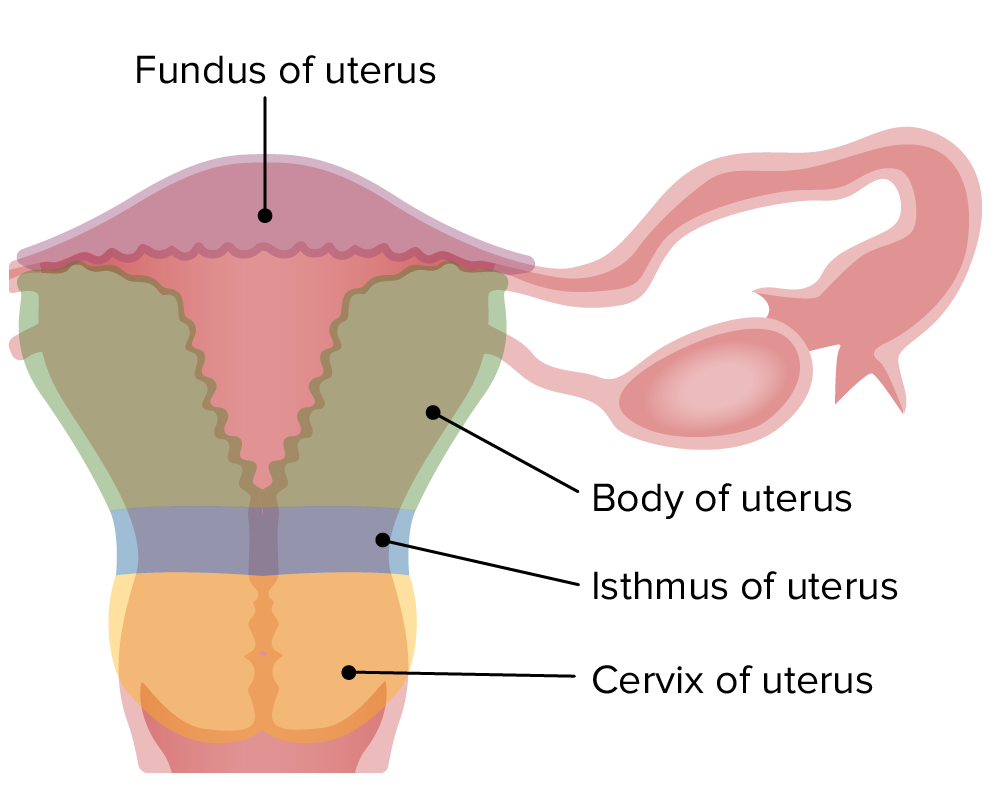
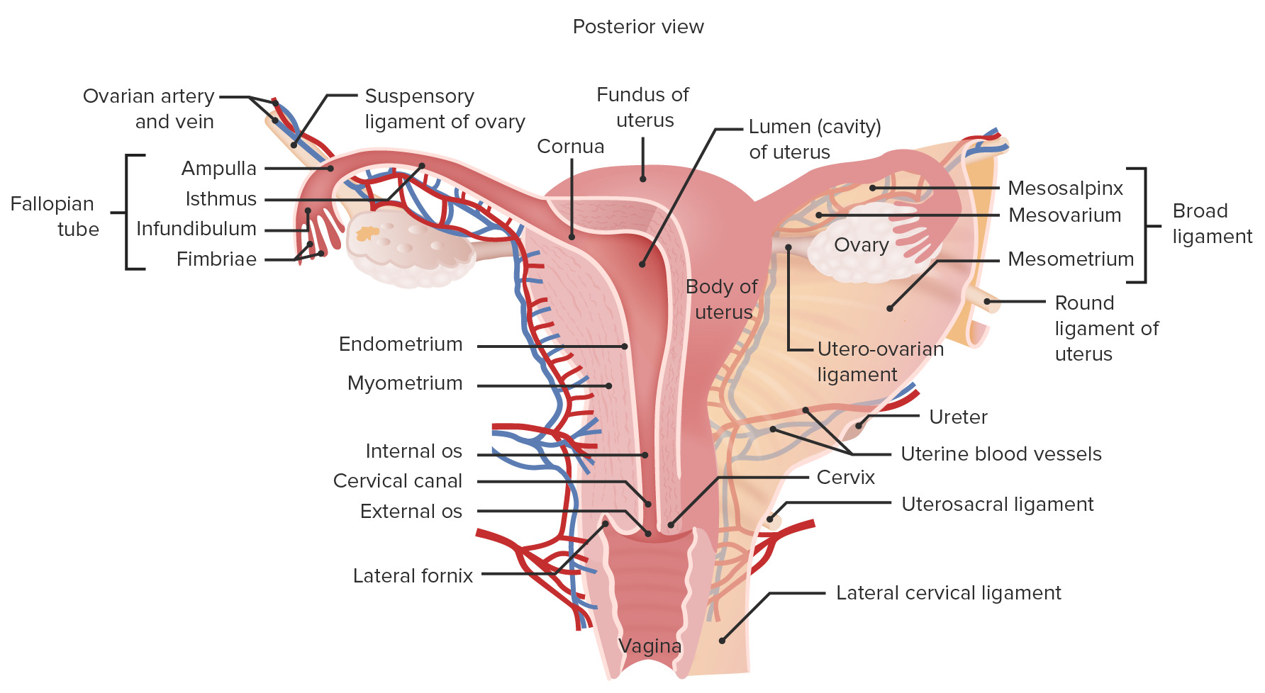
00:00 Here is a diagram and also a section through the cortex of the ovary. I want to briefly explain this diagram and this section, and then I’m going to move on and explain the various stages of follicular development. The central figure labelled A here summarizes what occurs in the ovary throughout the ovarian cycle. It shows a series of very round circular structures, follicles undergoing various stages of development during the ovarian cycle. The ovarian cycle is divided into three phases. The majority of the phases you see in this diagram A is follicular phase where follicles undergo growth. They get bigger. And those growth changes, you see on the right component of the diagram labelled B. These follicles go through a growth phase where they do a number of different things structurally. And I’ll show you these sorts of structures as we go through this lecture. But there's a couple of main points to remember as we go through. 01:20 And I think they’re illustrated fairly well on the right-hand side of the diagram, figure B. Follicles go through stages where they develop an epithelial layer around the oocyte. 01:37 That epithelial layer is called the follicular layer, initially. And then as the follicle goes through a phase of growth, that layer becomes very multilayered or stratified, and they’re called then the granulosa layer which I’ll talk to you about later on. 01:56 So that’s one event that happens structurally as these follicles develop during the follicular phase of the cycle. The oocyte also increases its size. It’s not shown that clearly here, but that’s another event that we need to recall as we go through this lecture. 02:16 On the outside of this growing follicle are two layers that form. They’re called thecal layers, an outside layer called the theca externa, and an internal layer called the theca interna. 02:31 The externa is merely a connective tissue capsule around the follicle, carrying blood vessels into the theca interna. The theca interna, we’ll see later, is a very vascular layer. It’s also very important because it produces precursors for the granulosa cells of the follicle to produce estrogens. And the third change you see when you look at follicular growth is about day six or day seven of the follicular phase of the cycle, suddenly a number of these follicles are stimulated to grow larger. They’re stimulated to proceed towards the process of ovulation. And what happens then is they develop an antrum or a space shown at the very base of the diagram on the right-hand side. It begins in a secondary follicle, and that antrum gets bigger and bigger. It accumulates with fluid. And the cells are pressed out on the outside, the granulosa cells. And that follicle is now a very large follicle capable of ovulation. But as we’ll find out, only one follicle normally ever ovulates. So, this is just the summary of follicular growth. One thing to bear in mind though is that the majority of follicles that undergo this growth development never get to ovulate. And at various stages of follicular growth, at any stage, follicles can often just degenerate, in fact, most of the time they do degenerate, a process we call atresia. Go back to the middle illustration diagram now. And those little follicles shown there in the diagram of the ovary, after ovulation, the ruptured follicle, the one that ovulates is converted into a corpus luteum. And that corpus luteum is going to produce progesterone. 04:44 The follicles, as they grow, produce estrogen. So on the bottom left-hand side, you can see a histological section through the ovary. And I’m going to now go through the stages of follicular growth that I’ve just summarized, but actually show you pictures or images taken from histological sections of each of these stages. And there are three major stages I’m going to show you. Firstly, the primordial follicle, and they are the follicles that are there at birth. They’re actually in the ovary prior to birth. And then I’m going to show you details of the growing follicles, the primary and the secondary follicles, and then finally, the mature or Graafian follicle. On the image of the slide on the bottom left-hand side, if you look very carefully in that histological section, you can actually see details of all those sorts of follicles. The very large circular follicle you see in the bottom left-hand corner of this section is that large secondary or mature follicle. You can see the antral space. 06:04 You can see the circular oocyte. And then if you look again at the section to the right of that large mature follicle, you can see another follicle. It’s a very well-developed primary follicle. There’s no antrum there. This could make out a very clear oocyte, or the very, very prominent nucleus, a nucleolus. And then the little tiny ones out in the cortex are going to be primordial follicles and very small primary follicles.
About the Lecture
The lecture Ovary: Stages of Follicular Development – Female Reproductive System by Geoffrey Meyer, PhD is from the course Reproductive Histology.
Included Quiz Questions
Which of the following is the main type of follicle that responds to follicle-stimulating hormone?
- Primordial
- Secondary
- Primary
- Postovulatory follicle
Which of the following types of cells form a multilayered cluster of cells surrounding the oocyte in the preovulatory follicle?
- Granulosa
- Mucosa
- Submucosa
- Antral
- Oocyte
What do the granulosa cells produce before ovulation?
- Estradiol
- Progesterone
- Growth hormone
- Androstenedione
- Testosterone
What is the main hormone produced by the corpus luteum?
- Progesterone
- Estrogen
- Testosterone
- Androstenedione
- Estradiol
The follicles present in the ovaries at birth are best referred to as which of the following?
- Primordial
- Primary
- Secondary
- Antral
- Neonatal
Author of lecture Ovary: Stages of Follicular Development – Female Reproductive System

Geoffrey Meyer, PhD
Customer reviews
5,0 of 5 stars
| 5 Stars |
|
5 |
| 4 Stars |
|
0 |
| 3 Stars |
|
0 |
| 2 Stars |
|
0 |
| 1 Star |
|
0 |
