Playlist
Show Playlist
Hide Playlist
Ovary: Corpus Luteum – Female Reproductive System

-
Slides Female reproductive system ovary.pdf
-
Reference List Histology.pdf
-
Download Lecture Overview
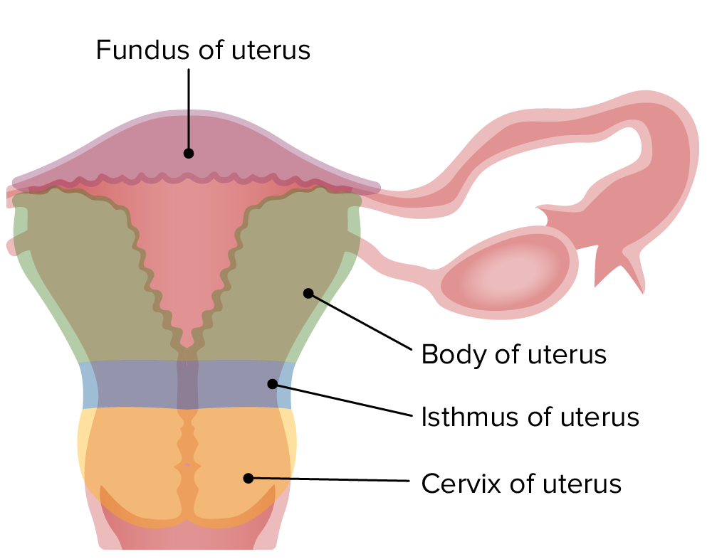
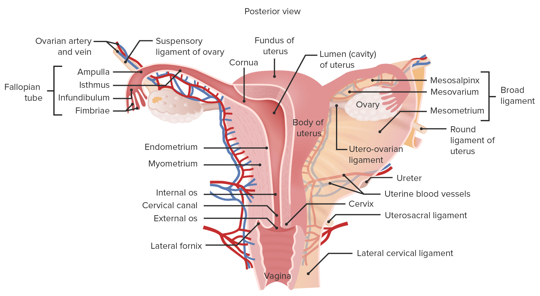
00:00 Now, here’s a corpus luteum. On the left-hand side, this is an image, a section through a corpus luteum from a recently ruptured ovarian follicle, Graafian follicle. All the red stained material you see is blood, because when the follicle ruptures, there’s some bleeding. And there's some growth blood vessels from the theca interna into the corpus luteum mass. Finally, that corpus luteum will grow to be a certain size. 00:35 And that antral space you see, which was previously part of the antral space of the follicle, will be converted into a very small fibrous core. And those blood vessels as they grow in, as I said earlier, establish an endocrine tissue, an endocrine organ. Whereby, the granulosa cells have changed structurally and they become vascularized, and they now have the capacity to produce progesterone under the influence of LH, which I've described earlier on. And this progesterone now coming out of the corpus luteum suppresses the development of follicles in the ovary during this luteal phase of the cycle. My Ph.D. was on the corpus luteum. 01:27 I looked at the growth of the corpus luteum, and I looked at how blood vessels grew into the corpus luteum, because one condition that prevents women from maintaining pregnancy beyond a few months is called luteal insufficiency. It’s the inability of the corpus luteum to produce progesterone. So my research project for my Ph.D. was actually looking at this structure. So I have a very soft affinity for it, an affection for the corpus luteum. 01:57 On the right-hand side, you can see a high magnification picture of a part of the corpus luteum. The bluey, purply stained cells represent the previous granulosa cells of the follicle. 02:16 We call them now granulosa luteum cells. The very paler stained cells you see, represent cells from the theca interna, that have invaded into the developing corpus luteum from the ruptured follicle. They’re called the theca lutein cells. So we have theca lutein cells and granulosa lutein cells. And the relationship between these two cell types is similar to what the relationship was like when they’re in the follicle. The granulosa lutein cells are now producing progesterone. They’re also producing estrogens because the theca lutein cells again provide those granulosa-luteal cells with the precursors for them to be out to secrete estrogen, just as it was in the follicle. Now, the corpus luteum just means yellow body, because when you look at the corpus luteum in fresh ovary tissue, it just looks yellow. And that’s because all those lutein cells have accumulated cholesterol stores, lipid droplets that is important as being a precursor for producing progesterone and for producing estrogen. So that’s what’s called the yellow body. When the corpus luteum has stopped functioning, it’s because it has its support withdrawn from the pituitary. 03:58 LH with support is withdrawn. And that happens about the sixth day after ovulation. Why does that happen? It happens normally because, after ovulation, we need to think a little bit about what happened to the oocyte. At the moment of ovulation or just prior to ovulation, the oocyte completes its first meiotic division, and enters its second meiotic division but is arrested. So at ovulation, the oocyte is quite large about 150 microns in diameter. 04:44 And you can see the second polar body alongside it, still enclosing the space between the oocyte and also the zona pellucida. And that’s a sign that ovulation has occurred. That oocyte, having entered the second meiotic division, is now called a secondary oocyte. And as I said before, it will only complete its second meiotic division after fertilization. 05:22 That oocyte is collected by extensions from the uterine tube that I’ll show you in a moment. 05:28 It’s almost like erectile tissue. It wraps around the ovary at the time of ovulation so that the ovulated oocyte passes into the uterine tube, and down the uterine tube for the chance of being fertilized. If that oocyte is fertilized, it spends another three of four days moving down the uterine tube until it finally implants in the uterine wall, in the endometrium. About six days after ovulation, during which time, this corpus luteum is producing more and more progesterone in response to LH. But if implantation doesn’t occur, that support is withdrawn. And it’s withdrawn because as soon as the fertilized egg implants in the endometrium, then the endometrial cells secrete human chorionic gonadotropin. 06:36 And that immediately signals to the pituitary that there is a pregnancy, there is implantation, and that, therefore, maintains the secretion of LH. And therefore, it maintains the lifespan and function of the corpus luteum for a number of weeks and produces increasing amounts of progesterone and estrogens that are so important for the maintenance of the pregnancy. 07:04 But if pregnancy does not occur, implantation does not occur, then the support is withdrawn, as I said a moment ago a few times, and therefore, the corpus luteum degenerates. And also, because the support is withdrawn, follicular growth starts again, and that’s the beginning of another part or another ovarian cycle. The corpus luteum then, after these granulosa cells start not producing progesterone, and as the theca lutein cells stop providing the precursors for producing estrogens, this corpus luteum starts to involute. These cells die off, and finally, are left with a scar called the corpus albicans, and that finally again diminishes in just a fibrous scar within the ovary.
About the Lecture
The lecture Ovary: Corpus Luteum – Female Reproductive System by Geoffrey Meyer, PhD is from the course Reproductive Histology.
Included Quiz Questions
Which of the following is INCORRECT?
- The zona pellucida remains intact in the ruptured atretic follicle.
- After rupture of the mature ovarian follicle, the theca interna cells differentiate into the theca lutein cells of the corpus luteum.
- Granulosa lutein cells continues to secrete progesterone after ovulation.
- The corpus albicans is the regressed form of the corpus luteum.
- Human chorionic gonadotropin is secreted by the syncytiotrophoblast.
Which of the following choices provides the correct sequence of terms of an ovarian follicle after ovulation during a normal menstrual cycle?
- Corpus hemorrhagicum, corpus luteum, corpus albicans
- Corpus luteum, corpus hemorrhagicum, corpus albicans
- Corpus albicans, corpus luteum, corpus hemorrhagicum
- Corpus hemorrhagicum, corpus albicans, corpus luteum,
- Corpus luteum, corpus albicans, corpus hemorrhagicum
Theca lutein cells and granulosa lutein cells originate from which of the following types of cells, respectively?
- Theca interna, follicular cells
- Theca externa, theca interna
- Theca interna, theca externa
- Theca externa, follicular cells
- Follicular cells, follicular cells
From which of the following does the corpus luteum develop?
- From the ovarian follicle following the release of a secondary oocyte which is halted in meiosis II.
- From the ovarian follicle following the release of a secondary oocyte that has completed meiosis II.
- From the ovarian follicle following a release of primary oocyte which has completed meiosis II.
- From the ovarian follicle following a release of primary oocyte which is halted in meiosis II.
- Any one of the options provided is possible.
Author of lecture Ovary: Corpus Luteum – Female Reproductive System

Geoffrey Meyer, PhD
Customer reviews
5,0 of 5 stars
| 5 Stars |
|
5 |
| 4 Stars |
|
0 |
| 3 Stars |
|
0 |
| 2 Stars |
|
0 |
| 1 Star |
|
0 |
