Playlist
Show Playlist
Hide Playlist
Nucleus, Nucleolus and Chromatin

-
Basic Histology 02.pdf
-
Reference List Histology.pdf
-
Download Lecture Overview
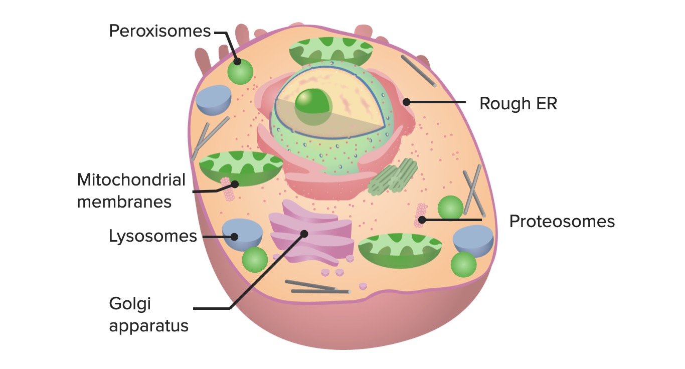
00:01 First of all, let me talk a little bit about the cell nucleus. It's shown here in diagrammatic form. 00:10 We normally talk about the nucleus during the interphase part of the cell cycle. 00:19 That's when the nucleus is bound by a nuclear envelope or a nuclear membrane and not undergoing mitosis. 00:31 The nucleus you see here has a tiny circular structure colored green, which is the nucleolus, so don't confuse the large outer circular structure that's the nucleus with the little structure in the middle, the nucleolus. 00:47 I'll talk about the nucleolus in a moment. 00:52 The normal cytoplasmic ratio, the ratio between the nucleus and the cytoplasm, is usually about 2:1 or 1:1 in most cells. 01:05 In cells that are immature or precursor cells such as lymphocytes that are yet to be told what to do really, then the cytoplasmic ratio, or at least the nucleus-cytoplasmic ratio can be about 1:1. Very little cytoplasm. 01:28 A lymphocyte, for instance, traveling around the body in the blood waiting to detect an antigen really isn't doing anything, but when it detects that antigen, it will then produce or grow its factory to create different components to help fight that antigen, And so the cytoplasm will increase in volume, but until then, the nucleus- cytoplasmic ratio is quite small. 01:59 Most cells that you will study in histology are mononucleate. 02:05 In other words, they only have one nucleus. 02:08 A couple of cells don't have a nucleus at all. 02:12 For instance, the erythrocyte or red blood cell or the thrombocyte or platelet. 02:20 Some cells actually have two nuclei or they're binucleate. 02:26 Some even can be multinucleated. 02:31 Have a look at this slide. 02:34 Look at the top left-hand image. 02:38 One of blood cells and right in the middle, you can see a neutrophil. 02:45 That neutrophil has what we call a segmented nucleus. 02:49 The nucleus is not a nice circular structure you normally would expect to see, but you can see it's divided into a number of little lobes. 02:57 It's that dark purple-stained structure within the neutrophil in blood. 03:04 On the top right-hand side, if you look at the very surface cells, they stain slightly more pinker, and if you look carefully at some of those cells, they each contain two nuclei or they're binucleate. 03:22 And down the bottom left hand side, this is a section through the heart, and if you look towards the top along the few cells that make up the top lining, there are a few cells there that look like they're two eyes looking out at you. 03:38 They're binucleate Purkinje cell fibers that help in the conducting mechanism through the heart. 03:46 Again, they're binucleate. 03:49 On the bottom right hand side, you see an image of two skeletal muscle fibers. They're multinucleated. 03:59 You can see very elongated nuclei running on the periphery of this skeletal muscle cell. 04:09 Now, I said skeletal muscle fiber earlier because really, when you think about skeletal muscles, those cells can extend long distances. 04:20 We have the sartorius muscle for instance in our thigh that can go from a region near the hip all the way down across the knee. 04:30 So these skeletal muscle cells traveling that distance, it makes more sense to call them a fiber. 04:38 But you can see how multinucleated they would need to be, and skeletal muscle fibers like you see here as well as the cardiac muscle fibers you see on the bottom left hand image, sometimes, when cells develop through the fusion of lots of smaller cells during development, we tend to call them a syncytium, so skeletal muscle really is a fiber that's a syncytium of lots of little cells that join together or fuse during the very early development of these skeletal muscle cells or fibers. 05:21 The shape of the nuclei is usually circular, but again as I might have mentioned earlier, they can be also elongated or columnar, and that's a good indication of the shape of the cell itself. 05:38 We've already seen in that neutrophil that the nucleus can sometimes be segmented, and in the case of a megakaryocyte, they can be multi-lobed. 05:51 Now let's look at them in a little bit more detail. 05:55 If you look at the nuclear membrane shown in the image here, you can see that it's a bilayer. 06:02 It's a double envelope. 06:06 We call it the nuclear envelope sometimes, instead of the nuclear membrane. 06:12 And what you can see in the left-hand side of the image is a whole lot of proteins that make up a nuclear pore, and look at the diagram on the right. 06:24 These nuclear pores occur all the way around the nuclear envelope, and they're very important because they allow the transfer of materials in and out of the nucleus. 06:38 For instance, RNA or ribonucleic acid needs to travel from the nucleolus out through these pores into the cell cytoplasm. 06:52 Focus again on this diagram. Again, pick out the nuclear pores that I just described and now, let's look inside the nucleus itself. 07:03 The most predominant structure you see is that large circular green-colored structure that represents the nucleolus, which makes ribosomal RNA. 07:17 The nucleoplasm occupies the remainder of the nucleus and includes a very fine scaffolding network of fibers within it and chromatin. 07:30 Chromatin consists of DNA strands and protein complexes. 07:36 If you look at the circular inset image down the bottom, it's a histological section showing you a section through the spinal cord and focusing on a large ventral horn motor neuron, that's that large structure you see towards the bottom, but the nucleus is that very clear circular structure in the middle. 08:01 The nucleolus is a very tiny, round, dark circle you see in the middle of the nucleus. 08:11 Let's focus now on the nucleolus. 08:14 Again, in the image, it's indicated by the colored green spherical structure, it has no limiting membrane. 08:30 Its job is to synthesize ribosomal RNA. It's basically at least 85% to 90% protein and the rest is RNA and DNA, all designed to synthesize ribosomal RNA which is essential for carrying out protein synthesis. 08:56 And again, that ribosomal RNA has to pass from the nucleolus, out of the nucleus, through the nuclear pores that I mentioned earlier. 09:08 And again on the inset, you see a section through the cerebellum in the brain, and the large dark-brown stained structure you see towards the top left and the bottom right of this circular inset, you can see within them these dark brown-stained cytoplasmic components, again, a lighter brown circular element which is the nucleus and then a little round dot which is the nucleolus and that nucleolus in those big cells is the same size as the nuclei of some very small cells in the background. 09:51 Those very small cells in the background are the granular cells of the granular layer of the cerebellum. 09:58 They're the smallest cells in the body. 10:00 They're around 4 µm to 5 µm in diameter so when you compare the size of those nuclei with the nucleolus in these big Purkinje cell fibers you see that I pointed out earlier, you can see that the nucleolus is roughly about 5 µm in diameter. 10:24 Again on the left hand side, you can see a large motor neuron with very purple-stained cytoplasm and again, this very pale-stained nucleus and the dark-stained circular structure, the nucleolus. 10:45 And on the right-hand side, you can see an electron micrograph image of the nucleus. 10:52 It's a little bit distorted in shape, but it shows you the very dark-stained nucleolus sitting in the nucleus and all that dark-stained material that you see within the nucleus is chromatin. 11:08 Some of it is around the outside appearing quite clumped, clumped chromatin whereas towards the center of this nucleus on the right-hand side, the chromatin is rather dispersed or lightest staining and that depicts the two sorts of chromatin we have in nuclei. 11:33 The lighter-stained chromatin that you would have seen on the previous slide is called euchromatin. 11:41 It's less compact. 11:43 The darker-stained chromatin that you saw on the periphery is called heterochromatin. 11:51 It's more tightly packed DNA. And on the right-hand side in the inset, you can see a close-up of that earlier picture I showed you of the kidney tubules, and if you're looking to the nuclei there, most of the chromatin is quite tightly packed, or most of it we would say would be heterochromatin. 12:17 Now, when it's less compact or when there's a lot of euchromatin inside the cell nucleus, it means that the chromatin is uncompacted, the DNA is unraveled, it's uncoiled, it's more open to transcriptional enzymes, it's more active and therefore, by looking at cells, you can sometimes depict various activity of cells particularly in relation to their production of proteins. 12:58 Have a look at this slide. 12:59 Here, most of the nuclei you see are dark-staining. 13:04 They've got lots of heterochromatin in them, so the cells aren't that active. 13:10 They're just sitting there waiting to be stimulated by perhaps a chemical messenger. 13:16 This happens to be a section in the tonsil where a lot of these cells are lymphocytes just waiting to be programmed to do something, so they're essentially inactive. 13:28 So their chromatin is very clumped. It's heterochromatin. 13:32 But cast your eye up to the top area of the image and you can see some rather larger nuclei. 13:42 They're more elongated. They're light-staining so they're depicting more euchromatin. 13:49 They're more dispersed chromatin, the chromatin that indicates that the cell is highly active, and those cells are actually fibroblasts in the early stages of laying down connective tissue elements within that tonsil that you can see. 14:07 Here again, just to summarize, chromatin on the left-hand side and the right-hand side, you can see the nucleolus and clumped heterochromatin and the lighter dispersed euchromatin.
About the Lecture
The lecture Nucleus, Nucleolus and Chromatin by Geoffrey Meyer, PhD is from the course The Mammalian Cell. It contains the following chapters:
- Nucleus
- Nucleolus
- Chromatin
Included Quiz Questions
Which of the following statements is INCORRECT?
- The nucleolus ultrastructure can be seen through a light microscope.
- The nucleolus has no limiting membrane.
- Protein-secreting cells may have more than 1 nucleolus.
- The nucleolus transcribes ribosomal RNA.
- The nucleolus is the largest structure in the nucleus of eukaryotic cells.
Author of lecture Nucleus, Nucleolus and Chromatin

Geoffrey Meyer, PhD
Customer reviews
5,0 of 5 stars
| 5 Stars |
|
2 |
| 4 Stars |
|
0 |
| 3 Stars |
|
0 |
| 2 Stars |
|
0 |
| 1 Star |
|
0 |
It was easy to learn and the speed was perfecy
Thanks... Dr Meyer very interesting and well presented information for rereading as a MD.
