Playlist
Show Playlist
Hide Playlist
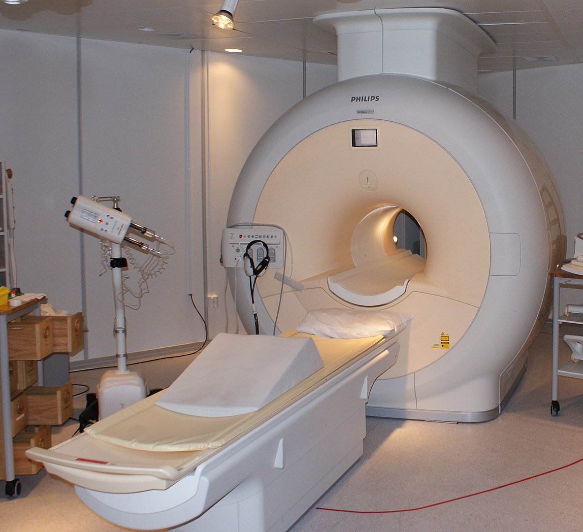
Magnetic Resonance Imaging (MRI)

-
Slides Intro Imaging MRI.pdf
-
Download Lecture Overview
00:01 So in this lecture we will be discussing the technical aspects of magnetic resonance imaging or MRI. 00:06 MRI images are created by the magnetic manipulation of hydrogen atoms. 00:12 This does not involve any ionizing radiation and MRI is actually very good for soft tissue abnormalities, fluid properties are what's used to create an image. 00:23 MRI is actually a very expensive examination and can take a significantly longer time than a CT scan does depending on the body part that's image, MRI images can take over an hour to perform. 00:34 MRI images are required using a superconducting magnet that has a continuously flowing electrical current that creates a permanent magnetic field. 00:44 The magnets contained transmitter coils, that transmit varying radiofrequency pulses that excites the protons to accentuate different tissue types. 00:53 The magnet also contains receiver coils that receive the signal back that's emitted by these excited protons and the information has then processed by a computer algorithm and that algorithm generates an image. 01:07 So the magnetic field strength of most MRI machines is commonly between about one and three Tesla, which is significantly higher than the Earth's magnetic field strength of about 50 microT. 01:17 Hydrogen Nuclei are very abundant within the body and they're composed of a single proton or a single positive charge so when the patient enters the MRI scanner all of these protons line a parallel or antiparallel to the magnet. 01:30 A short electromagnetic pulse is then transmitted and this pulse is called a radiofrequency pulse. 01:36 Coils that are located within the magnet are responsible for both transmitting and receiving an RF pulse. 01:43 And the frequency of that pulse changes the orientation of the protons. 01:47 Once the pulse is turned off the protons then realign with the magnetic field and release energy in the form of a radiofrequency pulse which is received by a receiver coils. 01:59 A dedicated computer then turns these pulses into an image that the radiologist can then view. The banging noise of the MRI machine which some of you may have heard is actually caused by vibration of the coils which is due to wrap an electrical pulses going back and forth. 02:13 So what exactly is T1 and T2? You may have heard people talking about, well, the T1 image shows this and the T2 image shows that, but what does that really mean? So T1 and T2 refer to the length of time it takes for the protons to recover or return back to their original alignment with the magnet from the time that the pulse was administered. 02:35 T1 refers to the relaxation time which it takes for the protons to align parallel to the magnetic field and T2 is the relaxation time for the protons to align perpendicular to the magnetic field. 02:48 So how are the T1 and T2 images created? Pulse sequences are predetermined imaging protocols that are specific to the body part that's being scanned and T1 and T2 are actually one of many different pulse sequences. 03:03 So T1 and T2 is based on TR and TE, so a short TR results in a T1 weighted image. 03:13 TR is the repetition time and that's the time between two radiofrequency pulses. 03:18 A long TE results in a T2 weighted image. 03:22 TE refers to the echo time and that's the time between the RF pulse and the echo that it creates. 03:28 So let's review MRI terminology. Again, every imaging modality has its own terminology and an MRI structures are referred to as hyperintense or hypointense which is different than CT in which the word densities used rather than intensity. So when a structure is hyperintense it means that it's bright and bright structures have more signal intensity and will appear more white. When a structure is hypointense it means that has low signal intensity and it's dark or will appear more black. 04:01 So what kind of structures appear bright on a T1 weighted image? Fat tends to appear very bright, which is also true for a T2 weighted image so fat generally appears relatively bright on both T1 and T2. 04:17 On a T1 weighted image proteinaceous fluid also appears bright. 04:21 On a T2 weighted image however simple fluid is what appears very bright. 04:26 Certain types of hemorrhage can actually appear bright on both T1 and T2 and hemorrhage can vary in appearance based on the age of the hemorrhage. 04:35 So this is an example of a T1 weighted image, you can see here this is a CT, this is an MRI through the brain and you can see that the cerebrospinal fluid is actually dark because this is a T1 weighted image and fluid is bright on the T2 weighted images. 04:52 You can see here that the skin on the scalp is actually a little bit bright and this is due to fat within the scalp. 05:01 This is an image through the abdomen and you can see that the liver is a little bit bright on the T1-weighted images but this area right here which actually represents a cyst within the kidney is dark because this is a T1 weighted image. 05:15 On T2 weighted images this image through the abdomen shows that the cyst that we saw is now very bright because it contains fluid and fluid is bright on the T2 weighted images. 05:28 This MRI scan through the head shows bright CSF or cerebrospinal fluid. 05:34 Again, because fluid will be bright on these T2 weighted images. 05:38 So on MRI signal can also be suppressed from certain types of tissues and this can be used for diagnostic purposes. 05:47 So signal is suppress so that it appears dark rather than bright in a situation where you would normally appear, you would you normally expected to appear bright. 05:55 So fat suppression is often use on contrast enhance images in order to better visualize the bright contrast. 06:01 So contrast will appear hyperintense and when you have a T1 weighted image, you want everything else to appear hypointense so that the bright contrast is easily seen. 06:11 So let's take a look at this. This is an example of a FLAIR image. 06:16 On this image you can see a T2 weighted image on the left and you can see that the cerebrospinal fluid is very bright. 06:25 On the right image here, which is the FLAIR image. 06:29 You can see that the cerebrospinal fluid is actually dark. 06:32 So this image is actually very similar, both are T2 weighted images. 06:37 However, on the FLAIR image the fluid is depressed. 06:40 So FLAIR actually stands for Fluid Attenuation Inversion Recovery. 06:44 And it's essentially a T2 weighted sequence in which the CSF fluid is suppressed. 06:49 So that other T2 hyperintensities are easier to see. 06:52 This is an example of when fat suppression would be useful. 06:57 So here we are looking at the pelvis and this is the region of the scrotum, you can see a scrotal mass in this patient. 07:04 This is actually an axial T1 image that demonstrates this heterogeneous scrotal mass. 07:10 You can tell that this is a T1 weighted image because the subcutaneous fat here is very bright. 07:16 This image as a similar image but has fat suppression. 07:21 So now you can see the subcutaneous tissue here which is fat containing is now dark and the mass is also dark indicating that it contains fat. 07:30 Here we have the two images side-by-side, we have the one, that is T1 weighted without the fat suppression and on this side we have the T1 fat suppressed image and you can see the differences between the two. 07:43 This image also has contrast with in it and you can see contrast within the vessels here and you can see a little focus of brightness right here, within the scrotal mass which represents contrast enhancement. 07:56 So MRI can be performed with or without contrast. 08:00 The contrast agent of choice for an MRI is Gadolinium. 08:03 It's a heavy metal ion that's chelated to different compounds in order to be used with MRI. 08:09 And most commonly it's injected intravenously but it can also be injected intra-articularly. 08:14 It's renally excreted and what it does is, it shortens the T1 relaxation time which results in structures appearing brighter on post-contrast T1 images than on the pre-contrast T1 images. 08:26 Nephrogenic systemic fibrosis is a very rare fibrotic disorder that affects the skin, joints and organs. 08:34 And it can actually occur in patients with renal failure who received Gadolinium. 08:38 Therefore, keeping this in mind, Gadolinium is actually contraindicated in patients with a GFR of less than 30. 08:57 MRI safety is very important. 08:59 It's very important to remember that the magnet is always on. 09:02 Ferromagnetic objects can become airborne and can overheat and cause a lot of injury and damage. 09:09 Ferromagnetic objects that are within a patient such as bullets or shrapnel can also move and cause injury. 09:14 So keeping this in mind, MRIs actually contraindicated in patients that have these objects in a location such as the eyes, where movement can cause injury. 09:23 And with mechanical or electrical devices such as cochlear implants, most pacemakers or drug and insulin infusion pumps. 09:31 I've actually heard of a case report where the cleaning person actually went into the MRI machine not realizing that this was an MRI machine and the entire cart got blown up and ended up within the MRI scanner destroying both the cart and the scanner. 09:43 So it's very important to keep this in mind. 09:46 An effort is actually being made for newer medical devices to be non-ferromagnetic. 09:50 So most joint replacements are non-ferromagnetic and a lot of new surgical and aneurysm clips are also non-ferromagnetic. 09:57 There's also a new pacemaker that's been developed that can go inside of an MRI machine. 10:02 So it's important to know which objects are ferromagnetic and which are not. 10:06 And you can often look up the actual device to see whether or not it's safe to be used within the MRI scanner. 10:13 Claustrophobia is a very common reason that an MRI scan is aborted. 10:17 Often pre-treatment with sedatives can be helpful and it's usually attempted and there are open MRI machines that are available that have a much larger or open bore. 10:26 However, they are of lower magnetic field strength which actually degrades image quality. 10:30 So again, you have to take a look at the pros and the cons, to see how important it is to perform the scan and if there's any kind of medication that the patient can be given to help them go into the machine, if they do have claustrophobia. 10:42 So let's summarize. 10:43 We've now reviewed radiography, CT, MRI and ultrasound. 10:47 These are the four most commonly used imaging modalities within radiology. 10:51 So radiography and CT both use ionizing radiation. 10:55 MRI uses electromagnetic pulses. 10:58 And ultrasound uses soundwaves or acoustic energy. 11:01 In terms of relative cost. 11:03 The cheapest ones are radiography and ultrasound. 11:05 Ultrasound being the most inexpensive. 11:08 The most expensive is actually MRI and CT falls somewhere in between. 11:13 In terms of portability, radiography and ultrasound are both portable. 11:17 CT and MRI require very large machines and are not portable. 11:22 Radiography and ultrasound can be performed within a few seconds. 11:26 CT is also relatively quick examination and can be perform usually within less than a minute. 11:31 MRI however, takes many minutes to sometimes over an hour to perform depending on the body part that's being imaged. 11:38 And contrast is used in both CT and MRI. 11:41 It may or may not be needed, depending on what's being evaluated. 11:44 However, in radiography and ultrasound there is no need for contrast.
About the Lecture
The lecture Magnetic Resonance Imaging (MRI) by Hetal Verma, MD is from the course Introduction to Imaging. It contains the following chapters:
- Definition of MRI
- What is T1 and T2?
- MRI Safety
Included Quiz Questions
Which of the following is NOT true regarding an MRI scan?
- MRI uses ionizing radiation.
- Magnetic manipulation of hydrogen ions creates images.
- MRI is great for soft tissue abnormalities.
- It is an expensive technique.
- It takes a longer time than a CT scan.
Which of the following regarding T1 and T2 is FALSE?
- T2 refers to the time it takes for the protons to relax and align parallel to the magnetic field.
- T2 is one of the many different pulse sequences, which are predetermined imaging protocols, and are specific to the body part being scanned.
- A long echo time results in the T2 image and is the time between a radiofrequency pulse and the echo it creates.
- A short TR (repetition time) results in a T1 image.
- T1 and T2 refer to the length of time it takes for the protons to recover or return to their original alignment.
Which MRI appearance of the following different body structures is correctly matched to the pulse sequence?
- A renal cyst appears bright on a T2 weighted image.
- CSF fluid appears bright on a T1 weighted image.
- A hepatic cyst appears bright on the T1 weighted image.
- Fat in the abdominal wall appears dark in T2 weighted images.
- A hemorrhagic pancreatic lesion appears dark on a T1 weighted image.
What differentiates Fluid Attenuation Inversion Recovery (FLAIR) images from T2 weighted images?
- In FLAIR, the CSF fluid appearance is suppressed.
- In FLAIR, the CSF fluid is brighter than in T2.
- In T2, hemorrhagic lesions are brighter.
- In FLAIR, the gyri appear brighter than the sulci.
- In T2, the cerebral cortex appears more enhanced.
Which contrast is an agent of choice for MRI?
- Gadolinium
- Barium sulfate
- Chromium
- Arsenic
- Yttrium
Which situation is suitable for MRI to be performed?
- A 21-year-old football player with a probable anterior cruciate ligament injury.
- A 60-year-old male with a probable diagnosis of avascular necrosis of the right femoral head and a history of pacemaker implantation.
- An 11-year-old male with a head injury and a history of cochlear implants.
- A 35-year-old male on an insulin pump, scheduled for MRI for a pancreatic lesion.
- A 45-year-old male with eye pain with a probable metallic splinter in the eye.
Author of lecture Magnetic Resonance Imaging (MRI)

Hetal Verma, MD
Customer reviews
3,2 of 5 stars
| 5 Stars |
|
2 |
| 4 Stars |
|
0 |
| 3 Stars |
|
1 |
| 2 Stars |
|
1 |
| 1 Star |
|
1 |
Slide 9 is problematic, specifically for the explanation of T2. "Perpendicular" is not a very good term. There is no aforementioned information that would help explain what T2 is that is given in this lecture. What should have been said if this was to be avoided would be, "Beyond the scope of this lecture".
Not very informative. She seems to be nervous and runs very quickly through the slides.
Excellent lectures. If you could add on some lectures on radiography for head and neck region and CBCT for head especially maxilla and mandible, that would have been great.
Explanation of the T1 & T2 Terminology and how it works is lacking. Could have used way more slides to visualize this: MRI in Practice by C. Westbrook et al show how this can be done in Chapter 1. Important concepts which need to be properly explained, in this video it looks like it has been half heartedly done.
