Playlist
Show Playlist
Hide Playlist
Macular Degeneration and Retinal Detachment

-
03 Retinal Disorders V2.pdf
-
Download Lecture Overview
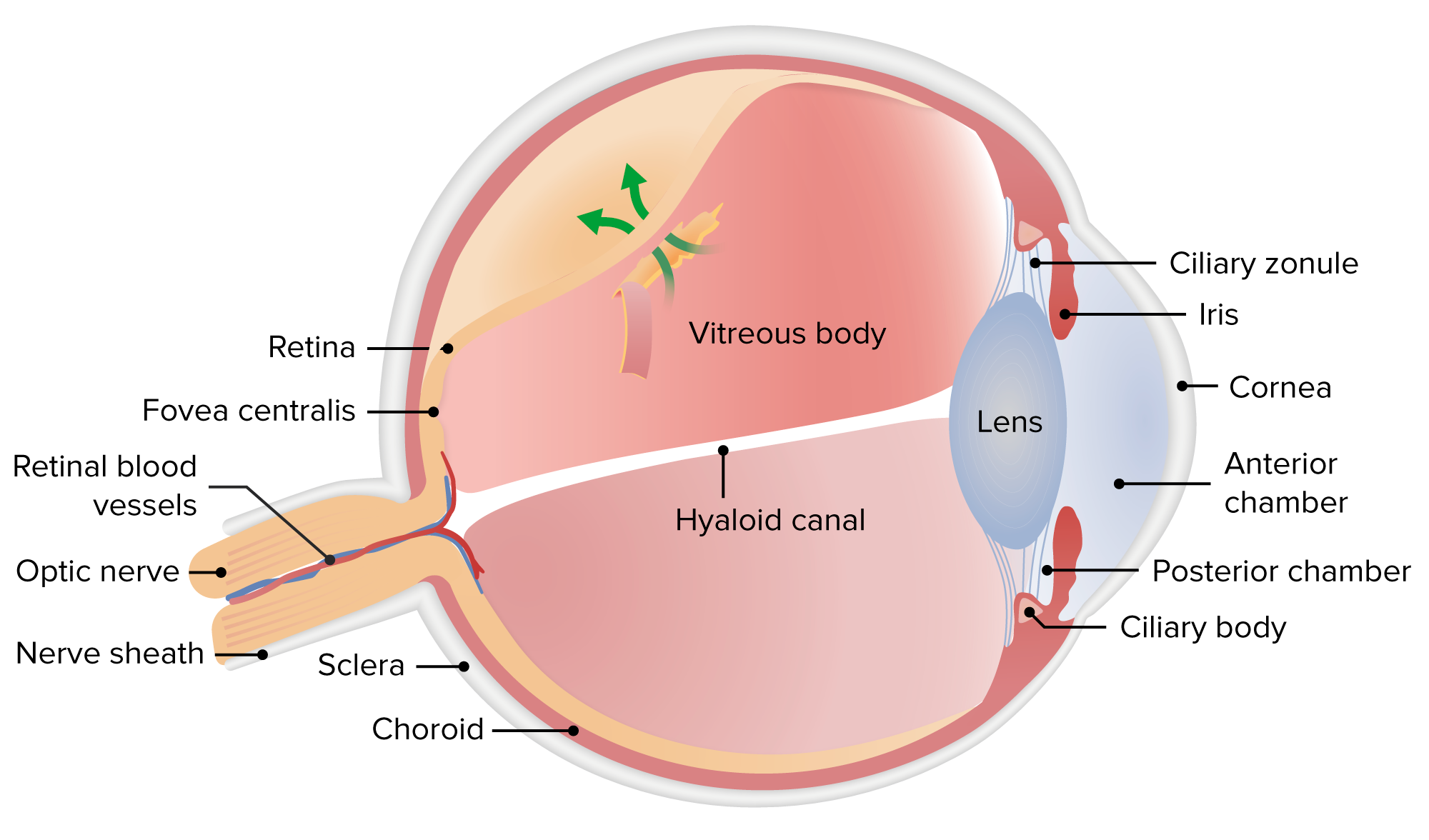
00:01 The macula. 00:02 Well the macula, do you remember, upon fundoscopic examination would be that area that has your cones and rods and photoreceptors. 00:10 And the area that has the highest number of them we then call the fovea. 00:15 This then provides, obviously, your visual acuity. 00:18 What happens when the macula undergoes degeneration? Maybe secondary to diabetes. 00:25 It is the number one cause of irreversible blindness in the US. 00:29 Number one cause. 00:31 90% of your patients thank goodness have what's known as dry which we then call nonexudative macular degeneration. 00:39 Why do I say thank goodness? Because the patient may not necessarily go into blindness as oppose to if you get exudative as we shall see. 00:47 10% of the cases could be wet. 00:50 In other words, exudative. 00:52 Exudative, of course, referring to leakage of fluid. 00:55 If it's the dry type, you're gonna have accumulation of this amorphous material. 01:01 Now, I need you to picture the retina. 01:04 I want you to move from the plexiform layers, outer, inner and then go into your photoreceptor. 01:11 And the photoreceptor layer is then laying upon your retinal pigment epithelium. 01:16 In between the retinal pigment epithelium in the photoreceptors right underneath there we have accumulation of a dry substance that's yellow and white and I'll show you a picture. 01:27 This is called drusen, okay? Drusen. 01:31 D, dry type of macular degeneration, nonexudative. 01:36 D, drusen. 01:37 That'll help you. 01:38 If it's exudative, this means that there's proliferation taking place. 01:43 So now you have what's known as neovascularization, abnormal blood flow growth therefore, contributing to a wet and exudative. 01:51 The waste product from photoreceptor cells is wet but drusen is often times referred to. 01:58 And the wet is due to the fact that you're leaking fluid such as blood and such. 02:03 And unfortunately, you're going to wet or proliferative. 02:07 It's just a matter of time before you go into irreversible blindness. 02:11 Rapid, rapid progression. 02:13 Hopefully, you've caught your patient before that ever happening through proper, proper education. 02:18 Let's manage the two types of macular degeneration. 02:22 If it's a nonexudative type, would you call this a dry or wet? Dry. 02:27 And accumulate what? Drusen. 02:31 D as in d. 02:32 And then accumulation of the waste product, the photoreceptors, between your photoreceptor and retinal pigment epithelium and your Bruch's membrane, remember that? Here the management will be antioxidant and zinc therapy. 02:48 In other words, multivitamins. 02:50 If you're going to exudative or the management of exudative which is a proliferative type, you need to make sure that you close this up in terms of your leakage. 02:59 So laser, photocoagulation and an anti-VEGF therapy drug. 03:04 For example, something like ranibizumab. 03:07 And therefore here, hopefully, you prevented the blindness and rapid progression from occurring. 03:13 On your left is the area that is extremely red and hemorrhaged. 03:19 We call this the wet neovascularization macular region is where we are. 03:24 So this is the exudative type, the wet. 03:28 And on the right what you see here on the far left is your optic disc. 03:32 In the right you have your macular region, but this time it doesn't appear as being hemorrhagic, does it? It doesn't appear as being red and wet. 03:40 If you do find these areas or plenty of little, little, little areas of white, white, white, white, white those would be the accumulation of that drusen that I've been talking to you about. 03:48 Once again, drusen will be for dry or nonproliferative macular degeneration and that drusen will then be located between the retinal pigment epithelium in Bruch's membrane. 03:59 Keep that in mind. 04:00 90% of your patient would have which type? The wet on the left or dry on the right? Dry on the right. 04:08 Age related. 04:09 Typical symptoms of macular degeneration are the distortion of straight lines and a central vision loss - this is evaluated with a grid test. To diagnose the specific type of M D you can use a fundoscopic exam, as seen on the previous slide. 04:25 Our topic here is retinal detachment. 04:30 Whenever do you might have neurovascularization, then what then might happen is that you are pulling the retinal layer off of the retinal pigment epithelium. 04:39 If that is occurring, then you have a fold that you're seeing here in the picture. 04:43 You see that area in the picture where it looks like a rainbow that's whitish? That represents the area of demarcation where retinal detachment is taking place. 04:51 Blindness, definitely something to worry about. 04:56 Hopefully you can get in there and do a little bit of photocoagulation so that you can savage as much of the vision as possible in your patient.
About the Lecture
The lecture Macular Degeneration and Retinal Detachment by Carlo Raj, MD is from the course Retinal Disorders.
Included Quiz Questions
Which of the following most commonly causes loss of central vision in the United States?
- Age-related macular degeneration
- Cataract
- Glaucoma
- Traumatic retinal detachment
- Diabetic retinopathy
It is believed that the failure of the eye to eliminate waste products from the cells of the rods and cones causes which of the following?
- Drusen
- Cotton wool spot
- Dot hemorrhage
- Flame hemorrhage
- Arteriovenous nicking
Drusen are found between the retinal pigment epithelium and which structure?
- Bruch membrane
- Choriocapillaris
- Choroid
- Outer limiting membrane
- Photoreceptor layer
Author of lecture Macular Degeneration and Retinal Detachment

Carlo Raj, MD
Customer reviews
5,0 of 5 stars
| 5 Stars |
|
5 |
| 4 Stars |
|
0 |
| 3 Stars |
|
0 |
| 2 Stars |
|
0 |
| 1 Star |
|
0 |
