Playlist
Show Playlist
Hide Playlist
Lymphatic Tissues and Organs

-
Slides 11 Human Organ Systems Meyer.pdf
-
Reference List Histology.pdf
-
Download Lecture Overview
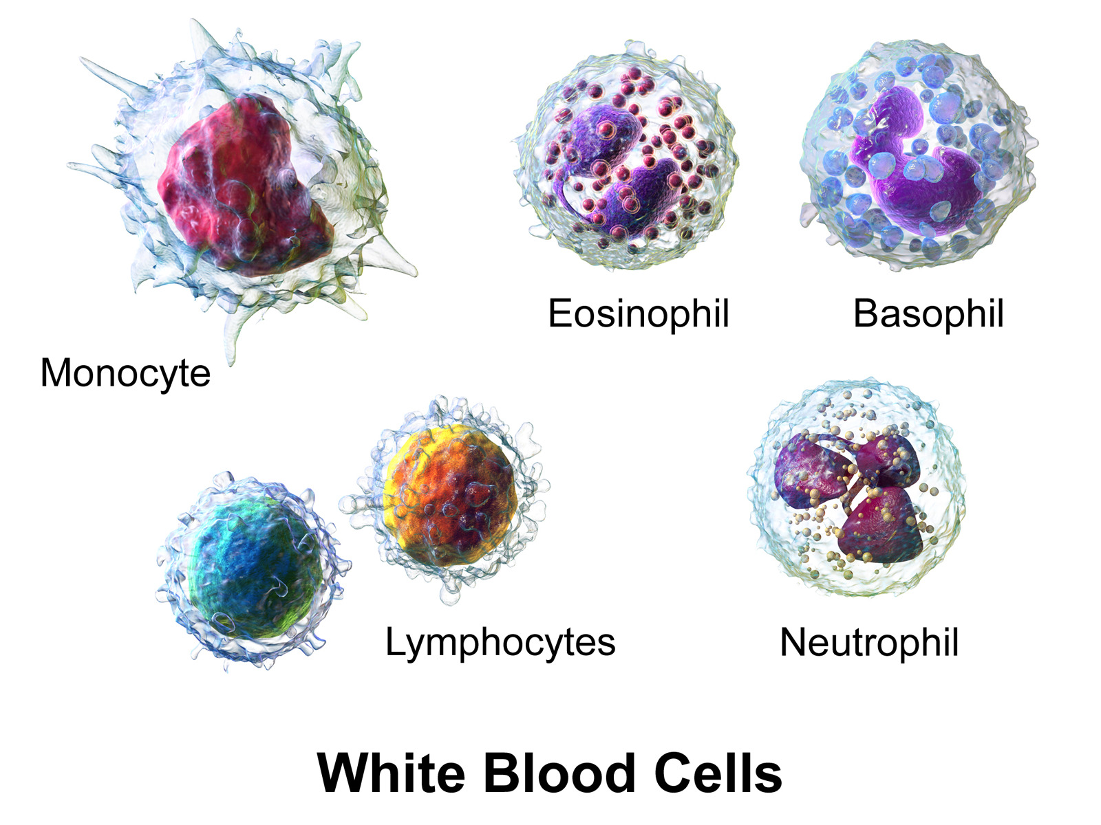
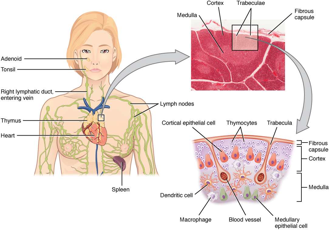
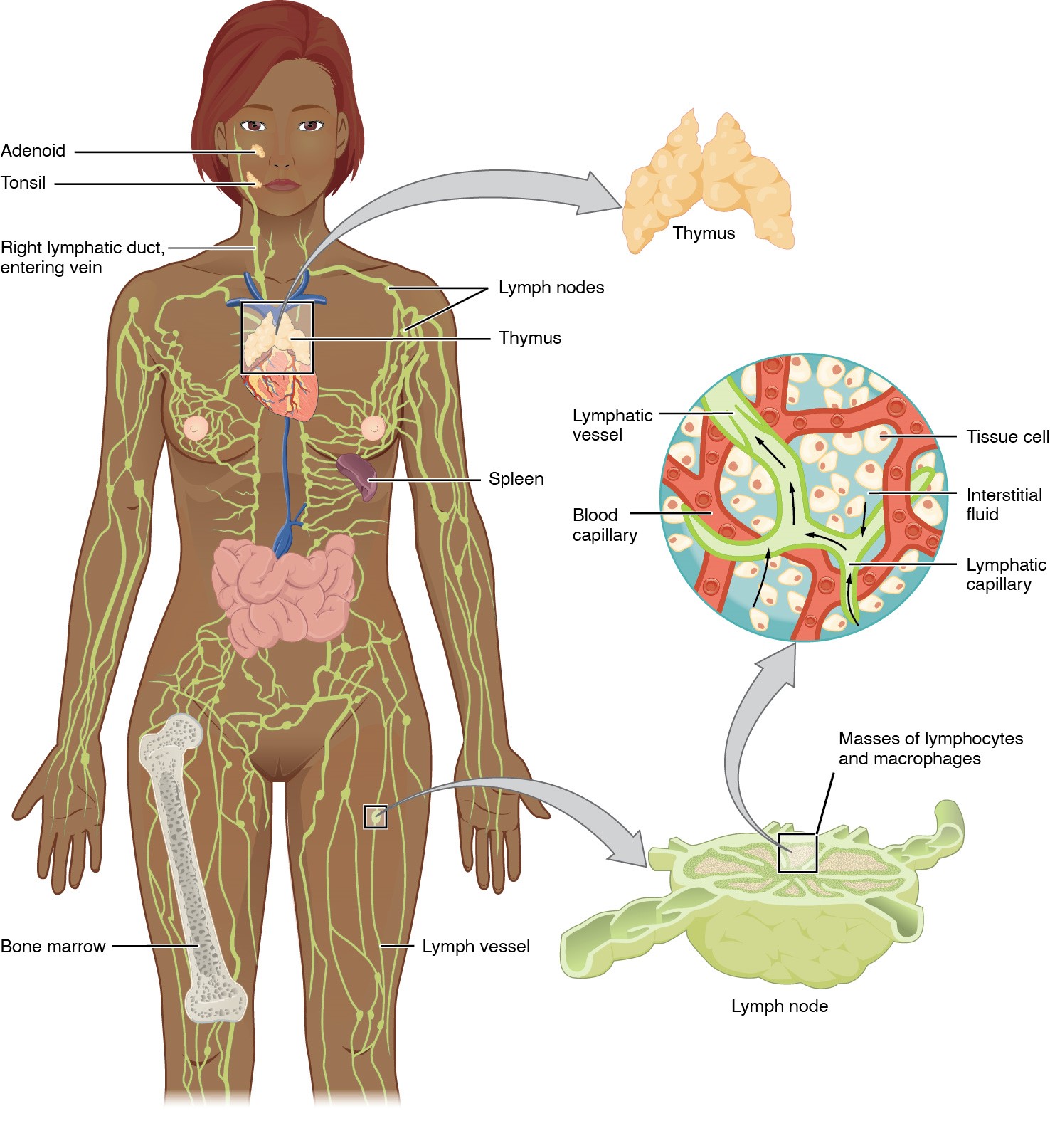
00:01 There are two major regions that the immune cells are first of all derived, and also where they’re located in most concentrations. 00:16 As I've mentioned earlier, the cells originally derive from the bone-marrow. That’s where they’re produced, or they can be produced in the gut-associated lymphoid tissues, or as I mentioned earlier, from the thymus. That’s where the T cells, remember, are educated. 00:34 These cells can then go and locate in the lymphoid organs that I’ll describe in this lecture. The nodules, the nodes, tonsil, and also the spleen. They’re all antigen-dependent, and they are the effectors of T and B cells. Once they’re switched on by an antigen, they become effector lymphocytes. And as we’ll see also, they remember so that they can mount a response a lot quicker to a secondary invasion of a particular antigen. So let’s now have a look at some of these lymphatic tissues, and also the lymphoid organs. Here are two pictures, two images taken from a part of the gut. One happens to be the stomach on the left-hand side, the stomach mucosa. The one on the right is taken from the small intestine, the duodenum. And on the left-hand side, you see what we call diffuse lymphatic tissue. 01:38 Lymphatic tissue is easily identified in mucosal surfaces because you see lots and lots of little tiny black dots. These represent lymphocytes. They don’t have much cytoplasm around them because they haven’t yet been activated. They’re waiting there on the surveillance duty. They are trying to recognize a particular antigen. And when they find that antigen that may have found its way across the surface of the epithelium, they recognize it, they bind to it, and then they can mount an immune response against it. On the right-hand side, you see evidence of that. When you see a nodule that you see on the right-hand side, a nodule is a pale staining germinal centre, and a darker staining corona, that represents a lymphatic nodule. 02:32 That is the morphological or the histological evidence that there is an immune response going on, because what’s happening there is that an antigen is passed across the surface. 02:46 It might have been ingested by an M cell or an enterocyte, a specialized enterocyte which is the M cell, and that might have ingested the antigen and then released it into the interstitial compartment underneath the epithelial surface to be recognized by a particular lymphocyte. 03:07 And when that lymphocyte recognizes that antigen, it binds to it, and it could immediately go into a situation where it can differentiate into a lymphoblast and then massively proliferate. 03:22 And that proliferation is evidence of that lighter staining germinal centre. And these cells can form memory cells or plasma cells. The plasma cells will secrete antibody. The memory cells are there to leave that area through part of the lymphatic system, a very small lymphatic channel, and then pass to a lymph node and beyond and get into the general circulation. 03:54 When they go to the lymph node, they can be detected and they can spark up another immune response and produce more memory cells and more plasma cells, and they in turn can then move and populate other mucosal surfaces just in case these invading antigens is coming across another surface. They can go out and be located for a long time sitting in these mucosal surfaces waiting for that antigen to come through. The corona you see there is really just where the peripheral cells are being pushed aside. They actually are lymphocytes that have detected the specific antigen that is being wrecked against in this situation. So they’re just like lymphocytes, as I’ve mentioned, that have been in the area. But as all this activity, this germinal centre gets bigger, they just get pushed to this side. Here is a tonsil. On the left-hand side, the tonsil sitting in the oral cavity is a perfect spot to detect any antigens that come in with the food. 05:02 They have tonsillar crypts. Tonsillar crypts are invaginations of the epithelial surface into the underlying connective tissue. And food debris gets caught in those crypts and it remains there for some time. And what happens is the lymphocytes, an antigen presenting cells, move across the epithelial surface into that debris, and then undergo surveillance to detect any antigens which they’re trying to detect. And if they do, again, they mount an immune response and you see a lymphatic nodule, as you see in this slide on the right-hand side. And again, these lymphocytes can always move away into lymph nodes nearby, create another lymphatic nodule, and again, proliferate more and more B cells, and more and more plasma cells, and again, more memory cells that can go and populate other surfaces as I described earlier. There is so much traffic across the tonsil surface, the epithelium of the tonsil that often, the epithelium is very, very difficult to recognize. It’s a stratified squamous epithelium, but as I said, in most areas where there’s so much traffic, it’s difficult to recognize that epithelial surface. You’ve seen on the left-hand side lymphatic nodules. 06:28 This is gut-associated lymphoid tissue. It’s very common in the ileum, part of the small intestine, combating against any antigens that found their way, passed all the usual defenses we often have. There are defenses we have where our first line of defense can be mucous, can be lysozymes. It can be the secretions on our skin surface, that's antimicrobial. 06:58 There's other ways in which we first try to prevent any antigen from getting into the body, the barriers. But if they do, then on the left-hand side, you can see a response against it. And on the right-hand side is the appendix, which is totally populated by lymphoid tissue. All those little dark dots are lymphocytes. And if you look very carefully in that slide, you can see nodules, again, reacting to antigens in the lumen of the appendix.
About the Lecture
The lecture Lymphatic Tissues and Organs by Geoffrey Meyer, PhD is from the course Lymphoid Histology.
Included Quiz Questions
Which of the following is NOT a secondary lymphoid organ?
- Bone marrow
- Lymphoid node
- Spleen
- Peyer patch
- Tonsil
Which of the following types of cells are the primary cells that proliferate and undergo diversification in the germinal centers of secondary lymphoid tissue?
- B cells
- T cells
- Monocytes
- Macrophages
- Natural killer cells
Which of the following types of cells become plasma cells?
- B cells
- T cells
- Monocytes
- Macrophages
- Natural killer cells
Which of the following statements regarding tonsils is INCORRECT?
- They are the main site for T-cell maturation.
- Their luminal surface is coated in non-keratinizing stratified squamous epithelium.
- They have antigen presenting cells on their surface.
- Antigens presented to B and T cells activate the adaptive immune response.
- They are secondary lymphoid organs.
Peyer patches are found in which of the following organs?
- Small intestine
- Thymus
- Spleen
- Lymph node
- Bone marrow
Author of lecture Lymphatic Tissues and Organs

Geoffrey Meyer, PhD
Customer reviews
3,0 of 5 stars
| 5 Stars |
|
2 |
| 4 Stars |
|
0 |
| 3 Stars |
|
0 |
| 2 Stars |
|
0 |
| 1 Star |
|
2 |
Not good didn't understand anything it should be in easy and simple wording
yup-side by side slide comparison so important bc histo is all in the details
This was a good general overview of the Lymphatic Tissue. Along with my textbook, this lecture was exactly what I needed.
He presents a list of the lymphocytes without the natural killer cells! Afterwards, he refers to them as if we know what these cells are. He also refers to lymphocytes as "key players", writing below a list of cells that we do not know in which category they belong. Busy slides, difficult language, not able to get his message across.
