Playlist
Show Playlist
Hide Playlist
Lymphangitis, Lymphedema and Other Disorders of Lymphatic Vessels
-
06 CVP Vein and Lymphatic Pathology.pdf
-
Reference List Pathology.pdf
-
Download Lecture Overview
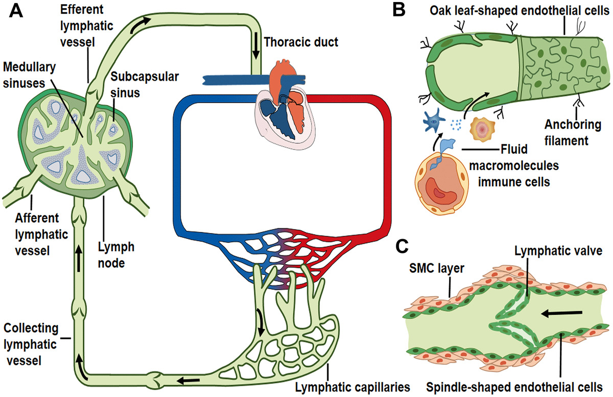
00:01 All right, that's vein pathology. 00:03 In very short order, let's do lymphatic pathology. 00:08 This is going to be a little bit quicker, so, pay attention. 00:10 Okay, so we have primary and secondary causes of lymphatic vessel obstruction. 00:16 Primary causes are extremely uncommon, but they're kind of interesting, so we'll talk about them briefly in a minute. 00:21 The secondary causes are much more common and that's because the lymphatics are the track by which we sample our various parts of our body, looking for infection. 00:32 Well, those infections may not just travel within the lymphatics, they may actually affect the lymphatics, you can have infections, so inflammatory processes, infectious processes and malignancy. 00:44 Again, malignancy will travel along lymphatics and lymph nodes. 00:48 So, lymphangitis is nothing more, nothing more than acute inflammation that's caused by bacterial seeding of the lymphatic vessels. 00:56 They appear as red painful subcutaneous streaks, you can actually see them, you can see them originating from a wound or some site of infection and you'll have inflammation all the way along there, should you choose to biopsy them. 01:09 It's often, not always, but often associated with an inflammation of the draining lymph nodes. 01:15 What's shown here in the green circle, is a tender enlarged lymph node, of a patient who had lymphangitis and then had lymphadenitis. 01:25 So, infection inflammation of the associated lymph node. 01:31 If the bacteria, are not contained within the lymph node, if we're not effectively dealing with the infection, they can pass into the venous circulation, recall that, lymphatics eventually make their way back up to the thoracic duct and that eventually dumps into the left subclavian vein, so, if we don't clear, it at one of these way stations then you can have, bacteremia and sepsis. 01:51 Okay, so those are kind of the secondary ones, and they're much more common. 01:55 Primary ones are kind of cool, so we'll talk about them briefly. 01:58 So, you can have isolated congenital defects, so, there are valves within the lymphatics and if those valves don't work, you can get a kind of a primary lymphedema. 02:11 There are familial syndromes called “Milroy disease” and there are others, that lead to defective milking of the lymphatics, it turns out that the lymphatics have a nice little milking activity, smooth muscle cells contracting all along their length and if that doesn't work appropriately, if it's not organized in a nice synchronized squeeze, then you can get lymphedema. 02:36 Secondary lymphedema can occur with tumors, obviously blocking flow, it could be internal or external, it can be surgical procedures that sever lymphatic connections and classically, when we have a woman with breast cancer, we will also take out these the axillary lymph nodes, associated with that breast and when we do that, we sever the lymphatics and then in the draining tissue, the arm, that would normally drain into those lymphatics, there's no place to go. 03:06 Post radiation fibrosis, similarly, will cause lack of flow through the lymphatics in the lymph nodes. 03:13 And then filariasis, so a parasitic infection, a certain worm, that has a peculiar predilection for lymphatics, and that will cause elephantiasis, that's the disease that you should remember, associated with filaria. 03:29 And then any inflammatory scarring, anything that will destroy, fibrose, compromise the normal lymphatic flow, will also give you secondary lymphedema. 03:42 So, what's happening here? So, we have some sort of lymphatic injury, lymphatic inflammation, there's increased hydrostatic pressure, because we're not able to drain fluid out of the tissue, that's associated with that, that gives rise to edema. 03:57 As we accumulate edema fluid in there, there’s going to be various fibrogenic mediators, that are going to lead to the deposition of extracellular matrix and we're going to get fibrosis. 04:09 Because of the edema, limiting flow into the tissue that will also compress venous structures, that have low pressure, and with the fibrosis, we're going to end up with inadequate tissue perfusion, and that will manifest the skin ulceration. 04:24 As we get accumulating edema and poor perfusion, we get what's called a brawny induration of the overlying skin, so, it becomes brown, and will also become quite edematous, and it can look like orange peel, so again, trigger alert next picture not really pleasant. 04:45 If the lymphatics dilate, they can rupture, you can get an accumulation outside of the lymphatics, of lymph that would normally drain. 04:54 If it was in the tissues you get edema, but in cavities we can get a chylous ascites, a chylothorax and a chylopericardium, so, this is just accumulation of lymph, in various tissues or various spaces, such as, the peritoneum, the thorax or the pericardium. 05:11 This is the image that I warned you about. 05:13 So, this is a very classic orange peel appearance, if you say it in French, it sounds better it sounds “Peau d'orange” which is, the orange peel. 05:24 And this is overlying a breast carcinoma, where the breast cancer has completely obstructed the lymphatics and we have this, kind of edema and involvement of the overlying skin. 05:39 This is a patient who had complete right sided opacity, shown here on chest x-ray, that is a chylothorax. 05:44 This is due to a rupture of a major lymphatic vessel, within the thorax, probably caused by malignancy. 05:53 And with that we've covered, in pretty quick order venus pathologies and lymphatic pathologies and hopefully, you've gotten some take-home messages to remember.
About the Lecture
The lecture Lymphangitis, Lymphedema and Other Disorders of Lymphatic Vessels by Richard Mitchell, MD, PhD is from the course Vein and Lymphatic Pathology.
Included Quiz Questions
Which of the following is a primary cause of lymphedema?
- Familial Milroy disease
- Tumors
- Surgical procedures
- Postradiation fibrosis
- Filariasis
What is the usual pathogenesis of peau d'orange of the breast?
- Blockage of cutaneous lymphatics by breast cancer
- Phlebitis
- Bacterial inflammation
- Arterial thrombosis
- Hypercoagulability
What is the underlying cause of filariasis?
- Parasite infection
- Bacterial infection
- Viral infection
- Fungal infection
- Autoimmune inflammation
Customer reviews
5,0 of 5 stars
| 5 Stars |
|
5 |
| 4 Stars |
|
0 |
| 3 Stars |
|
0 |
| 2 Stars |
|
0 |
| 1 Star |
|
0 |


