Playlist
Show Playlist
Hide Playlist
Lymph Nodes: Structure (Part 2)

-
Slides 11 Human Organ Systems Meyer.pdf
-
Reference List Histology.pdf
-
Download Lecture Overview
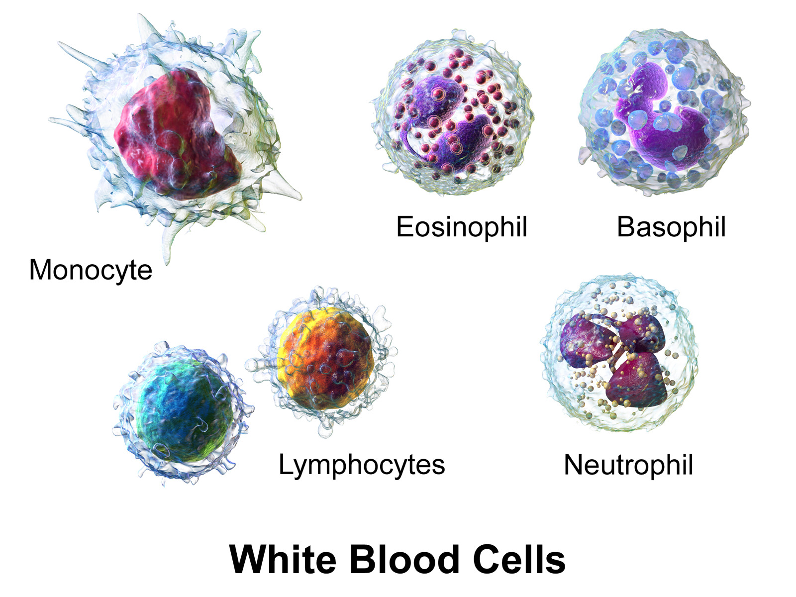
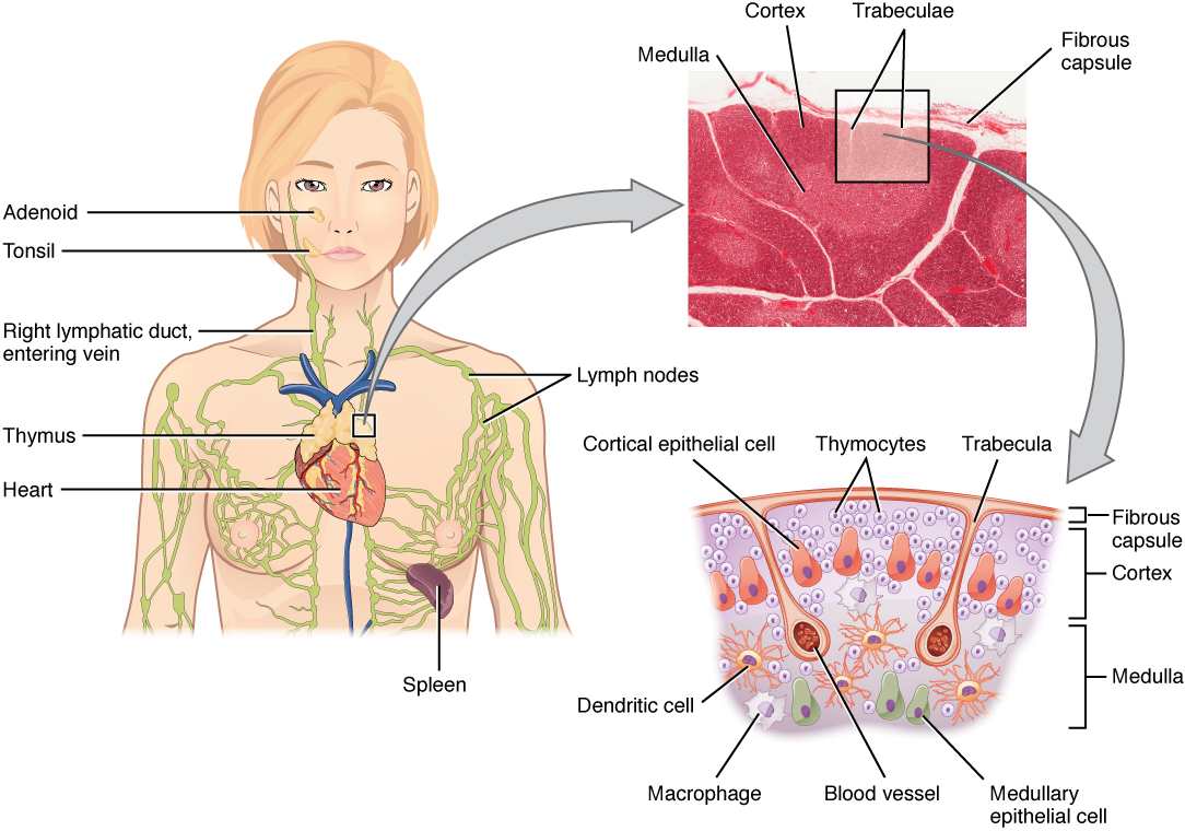
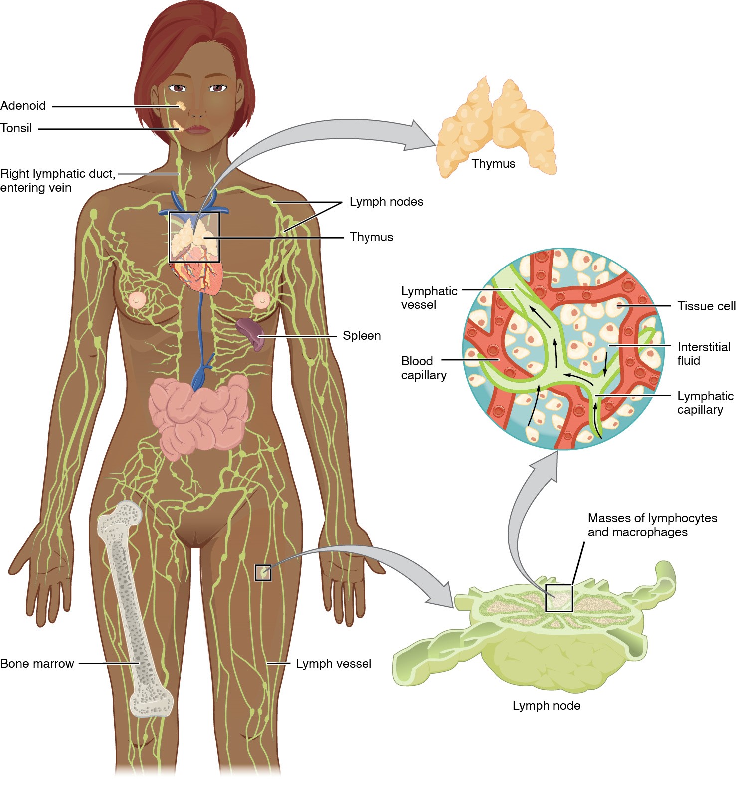
00:01 Here is a section of the lymph node taken at slight higher magnification. You can see the cortex labelled, the paracortex, the medulla, and the hilum. The medulla region contains lots of little tiny circular profiles, medullary rays of lymphocytes. And I’m going to mention those in a moment. The medulla is where the bulk of the lymph is going to drain through, on its way out from coming in from efferent lymphatics in the cortex. And that all drained into vessels and leave via the hilum shown there. B lymphocytes live in the cortical region. You can see a number of little nodules appearing there indicating these B cells there undergoing proliferation and differentiation into memory cells and plasma cells secreting antibodies. The paracortex is a region that is occupied primarily by T cells. So there is this difference in regional location of the B and the T cells. And we’ll also see that in the spleen, and I’ll point that out to you later on when we cover that organ. On this section, you can see a part of the cortex on the left-hand side. And there you can see, what I described before, little tiny lymphocytes in the vessel lumen. The efferent lymphatic vessels are coming in through the cortical region through the capsule of the cortex. And those little dark dots you see are lymphocytes packed in the lumen. On the right-hand side, you see a similar story. 01:59 You see the large efferent lymphatic vessel, and that also contains lymphocytes. 02:04 Those lymphocytes have probably come from other lymph nodes through the efferent one shown there, percolated through the lymph node. Others have come in through those venules I described earlier, circulate through the lymph node, and now they’re on their way out to finally go back into the vascular system, the blood. On the left-hand side here, you see part of the capsule. That capsule sends trabeculae of connective tissue into the lymph node. Just underneath the capsule there, you can see just a very clear space. And also near the trabeculum, that’s the beginnings of the sinus, the sinus spaces that contain the lymph. The lymph flows through those spaces, and then through all this network. The network you see in higher magnification on the right-hand side, you can see, if you look very carefully a reticular cell, you just see its nucleus wrapping around reticular fibres or collagen type III fibres which they produce. The collage type III fibres here are very well stained by the dark little lines you see indicating those fibres. Well, that’s that framework that I explained earlier. All these lymphocytes are percolating through that framework, and so to our antigens, and then they’re detected and dealt with. Now, the reticular sides are also very important because they attract, as I mentioned earlier, dendritic cells, macrophages, and other accessory cells. When they attract dendritic cells, they attract those dendritic cells specifically in the paracortex region. And there, those dendritic cells attract T cells. So that’s why there’s this localization of T cells in the paracortex mainly in the lymph node. On the left-hand side of this slide, you can see an image taken there at the cortex. You can see a lymphatic nodule. You can see the paracortex dominated by T cells. And then when the lymphatic nodule proliferates when the cells leave that nodule as B memory cells or plasma cells, they follow a little pathway down which is evident by the medullary cord you see on the right-hand side. The right-hand image is taken through the medullary region of the lymph node. And these long tiles of migrating lymphocytes go along these medullary cords. The plasma cells secrete antibodies. And finally, all these cells, all these lymphocytes coming from the germinal centre could leave the lymph node via the lymph, and then populate the rest of the body. This is a high magnification picture taken through the germinal centre of a lymph nodule. On the left-hand image, you can see a large lymphoblast. These are cells that have been reverted back from a stimulated lymphocyte and will undergo a series of mitosis or proliferation to give rise to cells that are going to form plasma cells and secrete antibodies, and also B memory cells which are going to populate other parts of the body so that they can recognize the antigen should've come across the surfaces at a later stage. On the right-hand section, there is an image showing you a follicular dendritic cell. These are dendritic cells. They extend long processes throughout the network. And again, this is the germinal centre. They also attract and bind antigen-antibody complexes and display them on the cell surface of these follicular dendritic cells. And then they interact with the developing B lymphocytes. And the B lymphocytes that can recognize these antigen immunoglobulin complexes on the cell surface and combine to that strongly survive. If you can’t bind to the surface of these antigen immunoglobulin complexes, as well as they should, then they go through a series of apoptosis because the follicular dendritic cell won’t support them. So these follicular dendritic cells are very important in training the B lymphocytes to be very specific for the immunoglobulin and antigen complexes on the surface. 06:56 And if they can recognize that complex very well, then that follicular dendritic cell saves that B cells from undergoing apoptosis and being digested by macrophages which also are located in the germinal centre to digest all the B cells that do not make the proper training and recognition of the antigens to which they are trained to recognize. Here is a section showing these high endothelial veins, high endothelial cell veins. They are cuboidal really. They’re not thin squamous like you see them in other capillaries. And they are the ones that put flags up and allow lymphocytes to attach to the endothelial cell, the lining of the capillary, and then move into the lymph node. So in a review, make sure you understand now the structure of the lymph node, how lymphocytes get into the node, the lymph nodule, and the region of the paracortex being specialized for T cells.
About the Lecture
The lecture Lymph Nodes: Structure (Part 2) by Geoffrey Meyer, PhD is from the course Lymphoid Histology.
Included Quiz Questions
Which of the following statements regarding the lymph node is INCORRECT?
- Follicular dendritic cells found in lymph nodes are derived from the bone-marrow hematopoietic stem cells.
- The germinal center is where lymphocytes respond to antigens.
- B cells are mainly found in the outer cortex.
- T cells are mainly found in the paracortex.
- Trabeculae are extensions of the capsule into the lymph node.
Which of the following statements is INCORRECT?
- Lymphocytes can enter a lymph node via efferent lymphatic channels and postcapillary high endothelial venules.
- Lymph nodes filter lymph.
- Follicular dendritic cells play an important role in supporting B lymphocytes during their differentiation.
- Macrophages are present in the lymph nodes and phagocytize lymphocytes deemed to be incompetent.
- Lymph flowing through lymph nodes can expose any antigen in the lymph to immune cells and initiate an immune response.
Which of the following is NOT a histological structure of the lymph node?
- Nucleus
- Cortex
- Paracortex
- Medulla
- Trabeculae
Where are B cells mainly located within the lymph node?
- Cortex
- Paracortex
- Medulla
- Hilum
- Capsule
Which of the following are important because they attract dendritic cells and macrophages?
- Reticular cells
- Capsular collagen
- Trabecular collagen
- Cortical tissue
- Medullary tissue
What is the main function of reticular fibers in the lymph node?
- Structural support
- Antigen presentation
- Phagocytosis
- B-cell differentiation
Which of the following cells within a lymph node specifically prevent the apoptosis of B cells through antigen presentation?
- Follicular dendritic cells
- Natural killer cells
- Macrophages
- Reticular cells
- Lymphoblasts
Author of lecture Lymph Nodes: Structure (Part 2)

Geoffrey Meyer, PhD
Customer reviews
5,0 of 5 stars
| 5 Stars |
|
2 |
| 4 Stars |
|
0 |
| 3 Stars |
|
0 |
| 2 Stars |
|
0 |
| 1 Star |
|
0 |
very clearly explained. perfect to complete your understanding about this topic
this lecture is very Comprehensive , concise and easy to follow
