Playlist
Show Playlist
Hide Playlist
Lymph Nodes: Structure (Part 1)

-
Slides 11 Human Organ Systems Meyer.pdf
-
Reference List Histology.pdf
-
Download Lecture Overview
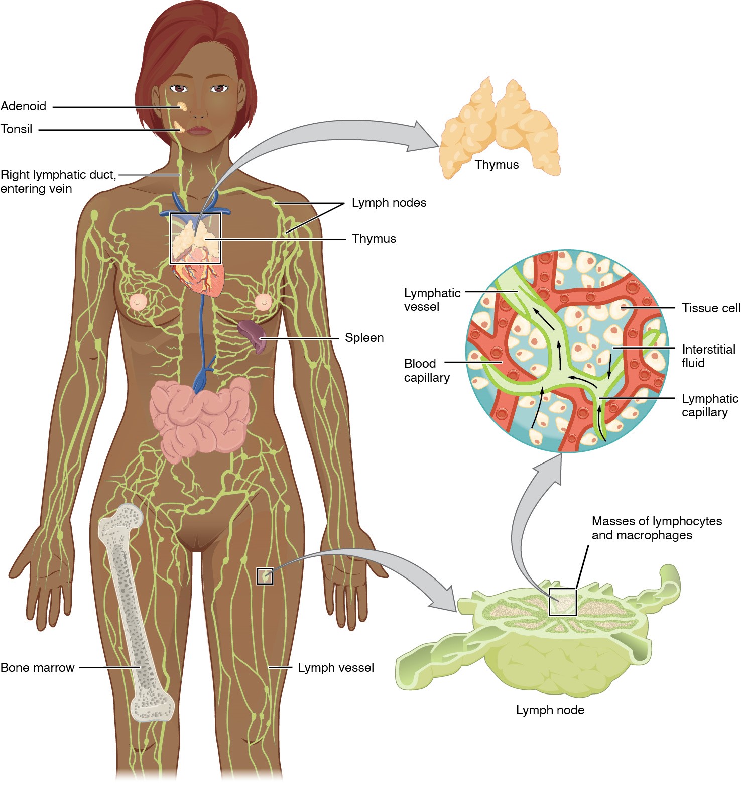
00:00 A lymph node is one of the basic components of the lymphatic tissue. It filters lymph. 00:08 On the left-hand side is a diagram illustrating the main structural features of a lymph node. 00:15 And on the right-hand side is a section through part of a lymph node. Lymph is formed from interstitial fluid in the peripheral tissues of the body. That lymph or that fluid has accumulated there. It's passed out of capillaries into the interstitium. And sometimes, that fluid is not then passed back into the postcapillary venules and returned into the vascular system. 00:45 There’s an accumulation of fluid in the interstitial space. Well, that excess of fluid is picked up by very fine little blind-ended lymphatic channels. These lymphatic channels communicate with each other. They join together to form a network and finally drain from tissues. 01:08 And they pass through lymph nodes. They pass through chains of lymph nodes, several lymph nodes before that lymph is then finally returned into the venous system through a large lymphatic duct. For instance, the right lymphatic duct empties all the lymph into the vein at the junction between the right internal jugular vein and the right subclavian vein. Now, in that interstitial fluid, there may be microbes, antigens, pathogens, foreign cells. And because these blind-ended lymphatic channels are very permeable, much more permeable than the blood capillaries, then these toxins, microbes, etc, can get into the lymph. 02:00 And therefore, the job of the lymph node is to actually clean that lymph to detect those antigens. On the left-hand diagram, let me just point out the main features of the lymph node. You can see a capsule on the far left-hand side. You can see lymphatic channels, these lymph vessels entering into the lymph node. They’re called afferent lymphatic channels. And those channels pass the lymph through the lymph node through sinuses. The lymph sort of percolates through the whole network of the lymph node with its antigens, with its toxins, and that lymph then finally passes out through the efferent lymphatic channel in the hilum of the lymph node. These lymph nodes are like little bean-shaped structures, little kidney-shaped structures. 02:57 And then that efferent lymphatic channel will pass on and become maybe an afferent lymphatic channel on the next lymph node. So there is this series of lymph nodes or lymph passes through as I’ve explained earlier in this lecture. Now besides that, there are, and it’s not described here in the diagram, this massive cobwebby meshwork of reticular cells and reticular fibres forming the framework of the lymph node. And as I mentioned at the start of the lecture, those reticular cells can hold up little flags and attract in all the different accessory, cells and T and B lymphocytes. And because they attract these cells into the lymph node, those cells could detect any of these antigens that are passing through the lymph, percolating through this meshwork, and finally, getting into the efferent channel. So that’s a strategically good location to identify and mount a response against these antigens. Also, there’s the blood supply shown here. You have an artery, small vessel coming into the hilum of the lymph node, and then forming a capillary network. 04:22 And then a postcapillary venule collects the blood from the capillary network and then passes out as a vein. That postcapillary network is very specialized. It’s specialized to allow lymphocytes to pass out of the blood into the lymph node. And this is how lymphocytes circulate. They go from the blood, pass these high endothelial cell venules, they are called, and they pass into the lymph, into the sinuses, into the spaces where the lymph is, and they circulate through the network, and they look for antigens that they are trying to detect. 05:07 Should they find an antigen that they can detect, they start up an immune response and develop a germinal centre and produce B cells, plasma cells, and antibody. T cells do a similar thing except they attach and ingest the antigens. They don’t produce any antibodies. So that circulation is very important. And that’s why lymphocytes get into the lymph. They move back into the lymph, into the efferent vessel, and they can return back to the blood through the channels that I've described earlier. So when you see these afferent lymphatic channels coming into the lymph node, you’ll see them full of little lymphocytes on their way back to the blood system. On the right-hand side, you just see a low power magnification of the lymph node. You can see the cortex on the very outside. There’s a little nodule there if you look very carefully. You can see the hilum of the lymph node, the lighter stained area, and I’ll show you more details of that in the next slide. Here is a section
About the Lecture
The lecture Lymph Nodes: Structure (Part 1) by Geoffrey Meyer, PhD is from the course Lymphoid Histology.
Included Quiz Questions
Lymph is initially and directly formed from which of the following?
- Interstitial fluid
- Arterial blood
- Venous blood
- Lymph nodes
- Bone marrow
Which vessel does the right lymphatic duct most commonly drain into?
- Right subclavian vein
- Right vertebral vein
- Left subclavian vein
- Left jugular vein
- Superior vena cava
Which of the following statements regarding the anatomy of the lymph node is MOST ACCURATE?
- Lymph enters the lymph node through afferent channels on the convex side of the node.
- Blood enters the lymph node through arteries on the convex side of the node.
- Blood exists the lymph node through veins on the convex side of the node.
- Lymph enters the lymph node through afferent channels in the lymph hilum.
- Blood enters the lymph node through a vein in the hilum of the lymph node.
Which of the following is not a structure of the lymph node?
- Kupffer cells
- Afferent vessel
- Efferent vessel
- Capsule
- Medulla
Lymph enters lymph nodes through which of the following?
- Afferent lymphatic vessels
- Efferent lymphatic vessels
- Thymus
- Efferent arterioles
Author of lecture Lymph Nodes: Structure (Part 1)

Geoffrey Meyer, PhD
Customer reviews
3,7 of 5 stars
| 5 Stars |
|
2 |
| 4 Stars |
|
0 |
| 3 Stars |
|
0 |
| 2 Stars |
|
0 |
| 1 Star |
|
1 |
Thanx for making me understand lots of things I did not know before. I appreciate it alot.
gostei muito das classes, boa didatica, recomendo muito. algun dia espero que tenha audio em portugues ou espanhol.
He talks 6 minutes straight without changing a slide! It would be helpful to have bullet points in order to understand step by step what he is going to cover. Very bad teacher.
