Playlist
Show Playlist
Hide Playlist
Lung Examination: General Observation of the Patient – Lung Disease

-
Slides 05 LungExamination Basic.pdf
-
Download Lecture Overview
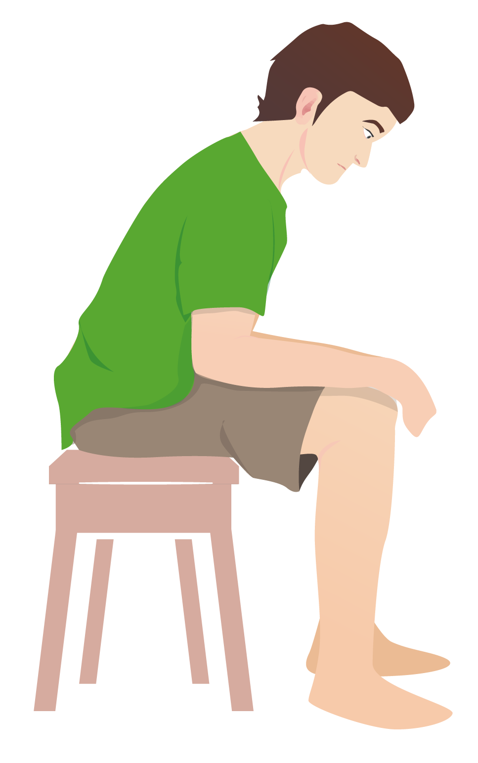
00:00 So general observation: does the patient look unwell? Do they have a fever? Patients with pneumonia and other severe infections, they look unwell. It’s obvious to anybody. It doesn’t have to be a doctor. It could be a lay person and they can tell that somebody is unwell. And the temperature needs to be measured. And if that’s high, then clearly they have a pyrexia and we need to think about what may be causing that pyrexia. 00:27 The level of consciousness surprisingly may be – is an important component of the respiratory examination, for acute presentations especially. Because in fact, when you have acute respiratory failure, the worse the respiratory failure the more likely you are to end up becoming drowsy. So reduced level of consciousness is a very bad sign for somebody who has severe respiratory failure in the acute situation. 00:58 Clearly for the respiratory examination, if the patient has difficulty in breathing when you’re examining them, that’s very important. And there are a few things we need to look for. 01:07 One is: what is the pattern of the breathing? And I’ll describe a few abnormal breathing patterns in the next couple of slides. 01:14 The second is: are they using accessory muscles? So, in the physiology lecture I described how ventilation is normally an active process on inspiration – requiring intercostal muscles and the diaphragm – and a passive process on expiration. But the skeletal muscles of the chest wall, the abdomen and the neck are not normally involved. But, when you have ventilatory disorders and you’re dyspnoeic and you have problems with severe breathlessness, then you start to use those muscles – the accessory muscles of breathing. So the sternocleidomastoids, the trapezius. A patient may lean forward and rest their arms so they can use their pectoralis major and minor to help with their respiration. And the abdominal muscles may be working very hard indeed. These are all evidence that the patient is struggling to breathe. 02:04 Pursed-lip breathing is an evidence of somebody actually having a problem with airways obstruction. 02:10 Pursed-lip breathing is when the patient breathes in and then breathes out against lips which are slightly closed, like that. That is a method of creating a little bit of end expiratory pressure as the patient breathes out. And the reason why that happens is that allows the airways which, otherwise, may collapse during expiration to be splinted open for slightly longer. And it’s a sign that patients with COPD have when they have bad airways obstruction. 02:42 Intercostal recession is where the muscles between the ribs sink in on inspiration and it’s a sign that the patient really is struggling with their breathing. 02:51 Another thing from the general examination is: are there any visible chest wall or general abnormalities? And I’ll discuss a few of those in subsequent slides. 03:00 So abnormal types of breathing patterns: the first of these is prolonged expiratory phase. 03:06 In normal respiration, inspiration is slightly longer than expiration. However, in airways disease, expiration is prolonged. So if you can – if you observe that a patient is taking longer over expiration than inspiration, that is a very early sign that they have airways disease in the examination process. 03:28 Cheyne-Stokes breathing is alternating slow and fast respiratory rate. The patient breathes fast, it slows down, it then stops and then it starts back up again. After a few seconds, it gets fast, slows down, stops for a few seconds. That’s actually a sign not of respiratory disease but normally of brainstem lesions or, perhaps, pulmonary oedema. When people have very severe pulmonary oedema, you see a Cheyne-Stokes breathing pattern at the very severe end of that disease. 03:59 Kussmaul is very fast, sighing respiration caused by a metabolic acidosis. So that’s where the low pH is driving a very fast respiratory rate rather than a respiratory problem. 04:12 Like conscious level, an irregular breathing pattern is a very bad sign. It means the patient is struggling badly with very severe acute respiratory problems and is likely to have a cardiorespiratory arrest at any minute. 04:25 Stridor is a wheeze on inspiration. It’s a very important sign because it identifies somebody who may have upper-airways disease. And those are a completely different type of disease to lower airways. It requires a different approach and can be, if it’s missed, fatal. 04:45 Excessive abdominal movements on inspiration occurs because of this, as we’ve already discusses, the issue about using your abdominal muscles to help with respiration when you’re struggling with your breathing. 04:56 Paradoxical abdominal movements is a very unusual situation and only occurs when you have significant diaphragmatic weakness. Normally on inspiration, the diaphragms flatten. Then the abdomen should go out. If the diaphragms are actually paralysed, as you breathe in using your intercostal muscles, the diaphragms come up a bit. And that will draw in the abdomen. 05:20 So, if on inspiration the abdomen sinks in, that’s called paradoxical breathing. And that’s a very strong indication there’s a respiratory muscle problem with the diaphragms. 05:31 So the visible abnormalities when examining the chest and observing the patient in general are: cachexia, marked loss of weight – we have a very thin-looking patient. That occurs in cancer, also in chronic infection and in fact it occurs in severe airways obstruction as well over time. Obesity is important because it does identify patients who are at risk of asthma because there’s an increased incidence of asthma in people who are obese, obstructive sleep apnoea and obesity hyperventilation. 06:01 If you have a single lung that has been shrunken either by previous surgery or previous scarring of some description, then that may be visible from the end of the bed because the chest wall will also be sunken slightly on that side. And you can see that normally at the top end of the chest where, if there’s a small lung, it will have a flatter aspect compared to the other side. And in fact when a patient takes a breath and you can see there will be less movement on the affected side as well. 06:29 Surgical scars are very important. A thoracotomy scar, obviously, might indicate somebody’s had some form of major surgery of the lungs. That could be a lung resection for whatever reason. There could be pleural-drain scars. These are very small, about that big. Normally in the axillary region. Quite easily missed but very relevant for people who may have had pleural disease in the past. Mediastinoscopy and mediastinotomy scars are surgical scars that people have had lymph-node sampling done. And the mediastinotomy scar will be here in the sternal notch. And a sternotomy scar would be just parallel to the sternum, somewhere around the third- or fourth-rib area. 07:04 We talked about hyperexpanded chest quite a lot in chest medicine. What we mean by that is people with airways disease – because expiration is a problem for them – they end up in fact increasing the volume of their lungs over time. So, on inspiration, they breathe in more air than they expire. Slightly different – slight gap in that volume. And, over time, that leaves the chest being hyperexpanded with the ribs more horizontal, the anteroposterior diameter being larger than it should be. And that’s visible in many patients with bad airways disease. 07:40 A kyphoscoliosis is where there is a curvature of the spine. A scoliosis is laterally, a kyphosis is anteriorly. And that indicates patients who may have problems with ventilation of the lung because of the abnormality of the curvature of the spine affects their skeletal movements during respiration and, therefore, ventilation. 08:00 Chest-wall masses are not common. Lipomas are pretty common but they’re obvious when you examine them that they are lipomas. But the ones that really matter are tumours that might be eroding through the chest wall. These are rare but, when they happen, clearly they are a very important sign. And there’ll be a firm mass than you can feel. And it’s rigid and fixed to the underlying chest wall. Not normally tender.
About the Lecture
The lecture Lung Examination: General Observation of the Patient – Lung Disease by Jeremy Brown, PhD, MRCP(UK), MBBS is from the course Introduction to the Respiratory System.
Included Quiz Questions
Which of the following breathing patterns matches the stated clinical situation?
- Kussmaul breathing and diabetic ketoacidosis
- Cheyne-Stokes breathing and exacerbation of asthma
- Stridor and severe COPD
- Paradoxical abdominal movements and pulmonary fibrosis
Leaning forward while breathing engages which muscle(s)?
- Pectoralis major and minor muscles
- Intercostal muscles
- Trapezius muscle
- Sternocleidomastoid muscle
- Diaphragm
Which of the following is indicated by pursed-lip breathing?
- Obstructive pulmonary disease
- Restrictive pulmonary disease
- Low Hb levels
- Connective tissue disease
- Muscular dystrophy
Cheyne-Stokes breathing occurs in which of the following conditions?
- Brain stem lesions
- COPD
- Cardiopulmonary arrest
- Metabolic acidosis
- Croup
Author of lecture Lung Examination: General Observation of the Patient – Lung Disease

Jeremy Brown, PhD, MRCP(UK), MBBS
Customer reviews
5,0 of 5 stars
| 5 Stars |
|
5 |
| 4 Stars |
|
0 |
| 3 Stars |
|
0 |
| 2 Stars |
|
0 |
| 1 Star |
|
0 |
