Playlist
Show Playlist
Hide Playlist
Lung Cancer Staging (TNM)


-
Slides 07 LungCancer Tumors and Metastases Neoplasias RespiratoryAdvanced.pdf
-
Download Lecture Overview
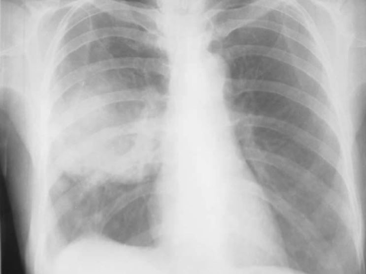
00:00 So staging of lung cancer is a little complex. This is a simplified table and there are three components to this. The first is the tumour size and simply enough the bigger the tumour the higher the stage and the higher stage is the ones which are less treatable. So for example tumours that is less than 3cm diameter is stage 1 on the tumour size staging, the T-staging, if it's 3-7 cm it is stage 2 T2. If it is above 7cms it's T3. However any of these tumours which are invading sites which you cannot resects such as the trachea large blood vessels of the spine and medially the tumour becomes stage T4. Nodal involvement is described either as none as in N-0, ipsilateral hilar nodes N1, ipsilateral mediastinal nodes N2, contralateral or extrathoracic nodes which means cervical lymph nodes, lymph nodes down in the gut, axillary lymph nodes and if they are involved it's an N3 disease. Metastases is very simple. Either the patient has an extra pulmonary metastases or it doesn’t. So M0- no metastases, M1- any metastases anywhere and it doesn't matter what size it is. It could be half a centimeter in the liver if that was metastases then that patient has M1 disease. This staging process is simplified a little to this and the reason for this simplification is that essentially, surgery is offered to patients with stages 1 & 2 but to not stage 3 or 4. So if somebody is presenting with mediastinal lymph nodes on the other side then they are not surgically operable. Anybody presenting with metastases is not operable. Ipsilateral mediastinal nodes it’s difficult. 01:58 They can be operated on some times but in general that probably means that’s not going to be operated on. Ipsilateral hilar nodes or just the mass in the lung can be operated on.
About the Lecture
The lecture Lung Cancer Staging (TNM) by Jeremy Brown, PhD, MRCP(UK), MBBS is from the course Lung Cancer.
Included Quiz Questions
Which of the following is NOT usually associated with small cell lung cancer?
- Hypercalcemia
- Hyponatremia
- Raised plasma cortisol level
- Eaton-Lambert syndrome
Which of the following statements is FALSE?
- A histological diagnosis is not possible for the majority of patients with lung cancer.
- Lung cancer can be diagnosed with a CT-guided biopsy of the metastasis of the adrenal gland.
- Sputum cytology is an insensitive test to diagnose a patient with lung cancer.
- Lung cancer is the most common cause of a CNS space-occupying lesion in patients over 65 years of age.
Which of the following is the highest stage of lung cancer?
- A 2-cm tumor that invades the spine
- A 7-cm lung tumor without involvement of the lymph nodes
- A 3-cm lung tumor with involvement of ipsilateral hilar lymphadenopathy
- A 5-cm lung tumor with ipsilateral hilar lymphadenopathy
- A 5-cm lung tumor without involvement of the lymph nodes
Which of the following statements regarding the staging of lung cancer is FALSE?
- Surgery is only offered to patients with tumors at stage 3 or higher.
- The stage of the tumor increases as the size of the tumor increases.
- TNM staging is the most common staging system used for staging lung cancer.
- Tumors with positive contralateral lung nodes are seen are usually inoperable.
Author of lecture Lung Cancer Staging (TNM)

Jeremy Brown, PhD, MRCP(UK), MBBS
Customer reviews
5,0 of 5 stars
| 5 Stars |
|
5 |
| 4 Stars |
|
0 |
| 3 Stars |
|
0 |
| 2 Stars |
|
0 |
| 1 Star |
|
0 |
