Playlist
Show Playlist
Hide Playlist
Lung Cancer and Tumors


-
Slides 07 LungCancer Tumors and Metastases Neoplasias RespiratoryAdvanced.pdf
-
Download Lecture Overview
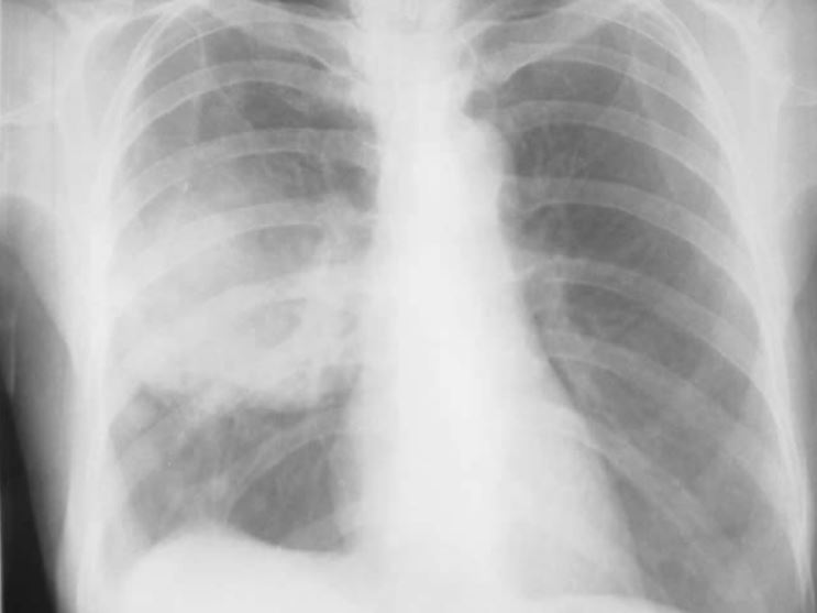
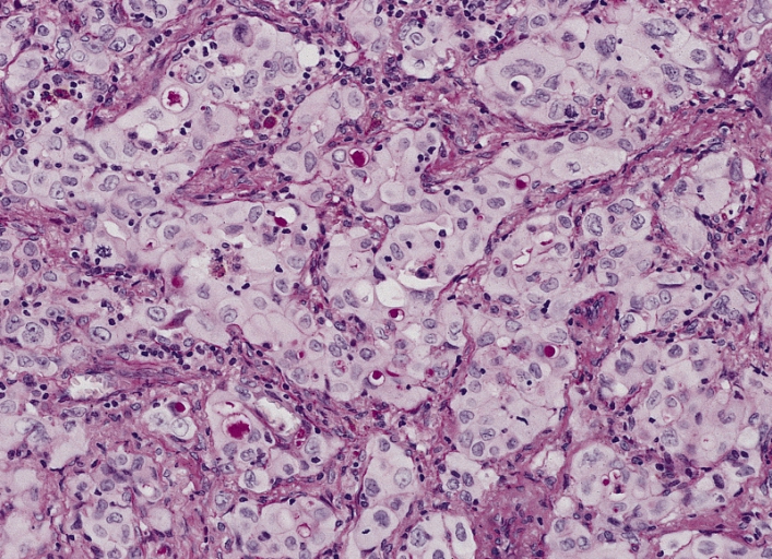
00:00 So the histology. This needs a biopsy and this is essential. Almost every patient presenting with potential lung cancer needs a biopsy to identify that there is a cancer and is now another more treatable condition and also to identify histology. So you can give the right medication. There are multiple different ways of biopsying. Patients who present with lung cancer are discussed those in a second. The principle is to use the least invasive, safest method and those tend to be the ones which are actually biopsies outside the lung. 00:35 So somebody presenting with lymph nodes metastases in the neck, actually there is a very easy biopsy to do. Because it could be done on ultrasound control and there is very little chance of any damage to any other tissue. Whereas somebody who has got a lung cancer which is right in the middle of the lung, the chance of biopsying that safely is pretty high. But you still got a 10% chance of getting pneumothorax or significant bleeding by doing a CT-guided biopsy of a lung tumour. The other principle is that you try to biopsy the highest stage lesion. So what I mean by that? Is there somebody presents you and they've got a chest x-ray and on that x-ray there is a mass. But when you do the CT scan it looks like they have a little metastases. The mass you want to biopsy is the little metastases. 01:21 The reason for that is that if you biopsy the liver and it turns out to be a lung cancer and that means they have stage 4 disease and they are incurable. Whereas you biopsy the lung you still have the question about what the liver mass might be. So that’s in that. 01:38 The other principle is to actually to target the higher stage lesion and that doesn’t mean you end up biopsying lesions outside the lung. Because it is safer and because that’s where the metastases may have been. Another important point is that the mediastinal lymph nodes are the break point of the many patients between whether they have curable disease or incurable disease. In this staging when the cancer has spread from the lung to the hilar lymph nodes it's still operable. When it spreads from the hilar lymph nodes to the mediastinal lymph node it becomes in operable. 02:11 So deciding whether the nodes are enlarged due to cancer is one of the important question that has to be answered in these patients. Because that decides whether they have curative therapy or not. Unfortunately the mediastinal nodes maybe enlarged in patients of lung cancer not just due to cancer involvement but due to other nonspecific information. That means that the fact they have a large mediastinal node on a x-ray does not mean that there is cancer involvement. What you have to have is a biopsy of that node to prove whether it is cancer or not. So there is a large range of biopsy methods that are used for identifying patients of lung cancer and confirming the diagnosis. 02:56 So for example, if you have a peripheral lung lesion, you normally need a CT guided biopsy through the skin by the radiologists. If it's a more proximal lesion, then actually those are normally reach by bronchoscopy. So you can do a bronchoscopy, you might be able to see the tumour as I showed earlier in the photograph of somebody with a right upper lobe lesion and you can biopsy it. You can get washings and that will give you direct answers to what's going on with it. If they have mediastinal lymph nodes as I have already mentioned that is a vital question whether they are involved with cancer or not and the way to identify with that involving cancer is to do a biopsy which is either under bronchoscope with ultrasound. So that is the endobronchial ultrasound guided biopsy where the needle is passed through the bronchial wall using the ultrasound to guide it into the mediastinal nodes that's been affected or it requires a surgical biopsy what we call as mediastinotomy or mediastinoscopy which is small surgical operations but still they do require surgery much more invasive than doing an EBUS guided lung biopsy. 04:04 Cervical nodes that I have mentioned could be biopsied using ultrasound control. Bone masses can be biopsied using CT guidance. Pleural metastases can be biopsied on the ultrasound or CT guidance. Adrenal masses and liver masses those can all be biopsied using radiological guidance. So these are the common methods by which the diagnosis may be made of lung cancer confirmed. Staging. So staging of lung cancer is a little
About the Lecture
The lecture Lung Cancer and Tumors by Jeremy Brown, PhD, MRCP(UK), MBBS is from the course Lung Cancer.
Included Quiz Questions
Which of the following statements regarding the principles of biopsy is FALSE?
- A biopsy must be taken from the lowest stage lesion accessible.
- A biopsy must be taken using the safest and least invasive methods.
- A biopsy of the mediastinal lymph nodes is useful for deciding if the patient's disease is curable.
- There are many different methods of obtaining a biopsy.
Which of the following techniques is usually utilized to sample mediastinal lymph nodes in patients with lung cancer?
- Endobronchial ultrasound-guided biopsy
- CT-guided percutaneous biopsy
- Transthoracic CT-guided biopsy
- Sputum cytology
- Bronchoalveolar lavage
Author of lecture Lung Cancer and Tumors

Jeremy Brown, PhD, MRCP(UK), MBBS
Customer reviews
5,0 of 5 stars
| 5 Stars |
|
5 |
| 4 Stars |
|
0 |
| 3 Stars |
|
0 |
| 2 Stars |
|
0 |
| 1 Star |
|
0 |
