Playlist
Show Playlist
Hide Playlist
Loop Spirometries – Laboratory Diagnostics
-
Slides Diagnostics PulmonaryFunctionTest RespiratoryPathology.pdf
-
Reference List Pathology.pdf
-
Download Lecture Overview
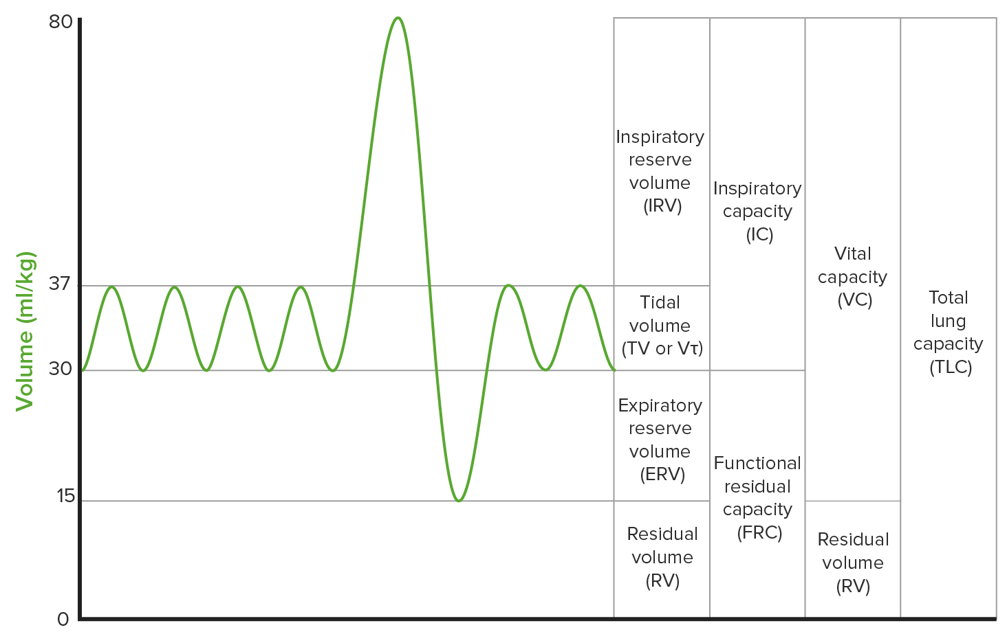
00:00 Let’s take a look at a important pulmonary function test and loop spirometries that are relevant to your clinical understanding. 00:07 Let’s take a look at the normal one first, right smack dab in the middle. Let's make sure, please, that we are absolutely confident about what the loops mean. What Y-axis, X-axis and the top and the bottom portion of the loop means. Let's begin. In the middle there, is normal. Next, well, what does that X-axis represent? Well for the most part, your volumes. I mean to say, well, how much is your lung filled with air? Let’s put it that way. You notice your residual volume and that will never change. You see the lines in blue, dashed line? That is residual volume, that is the air that is always going to be present normally in your lung, has to be. 00:52 Next, well, as you move in the middle from 0, 2, 4, 6, please understand, that is, take a look at the end on your right, the X-axis is labelled as volume. Keep it simple. So as you move from right to left, from 0 to 6, that obviously means an increase in volume. 01:12 Hmm, interesting! So, which one of these. which half of this loop represents inspiration? Please, please understand, it’s the bottom loop entirely. The entire bottom loop, from the beginning where it’s between 0 and 2 moved all the way up to 6, represents inspiration only. All of it. Stop. So now, you have ended at TLC, total lung capacity. That is full. 01:45 Okay. Now, where do you gonna begin? Well, let’s go through some physiology. Where is my diaphragm? Oh, it’s passively moving up. Good. Passively moving up. Now, what happens? Oh, the alveolar pressure is becoming positive. What does that mean? It’s going to squeeze the air out through the alveolar ducts, moving it out. Good. What happens to your pleural pressure? The pleural pressure is becoming less negative, isn’t it? So, maybe it was at negative 8, now it’s moving down from negative 8 to negative 7, negative 6, negative 5 and then you have the recoil or elastic pressure, right? And that elastic pressure is becoming well, equivalent. Equivalent to your pleural pressure. It has to. So, this is the process of expiration that you should be oh so comfortable with before we move on to the exhalation aspect of this curve. 02:43 There are three different things that are going on. There are three different pressures and please understand, at the end of expiration, we have now once again reached FRC technically. 02:54 At the end of inspiration, we have now technically reached FRC. That means now the alveoli is filled. Where am I? At 6. You picturing this? Alveoli is filled with air and its pressure is pretty much equivalent. It’s at 0. FRC. The pleural pressure and the recoil pressure are both equal at approximately 8, shall we say. They are going to begin the process of expiration. Here comes the air out. The entire top loop, ladies and gentleman, represents expiration. You will divide this into the first half of expiration upon the top loop and the second half of expiration, exhalation. Now, during exhalation, the second half, wow, there isn’t as much air than coming out. Hmm, what does that mean? Think about that. 03:49 As the air is coming out, then there is more pressure for it to close. So therefore, this is called dynamic airway expression. So that dynamic airway type of expression takes place usually the latter half of exhalation. This then brings us back to, well, where we started at? And you will notice here ladies and gentleman, that there is still little bit of volume left and that is your residual volume. This is a perfectly normal loop spirometry. 04:17 Let's go to our first pathology. You will find that the pulmonary function test in FEV1 to FVC ratio is less than 0.7 or 0.8 and it’s at 0.3. This to you without a doubt means obstructive. No doubt, no doubt. What you need to confirm that it’s only COPD or that it’s only obstructive? The volumes, right? So, they will tell you that the volume, meaning the TLC is increased or they show you loop spirometry here. So, on your left is obstructive. 04:50 How can you confirm that? Well, for two reasons. 04:52 Number 1, the red loop represents the normal loop spirometry. What’s inspiration again? The bottom half of the loop. Inspiration has taken place, it is leaves you to 6. I told you with that type of pulmonary function test that you have a ratio that is less than 0.3 or its at 0.3. Then where is the air trapped? In your lung. Think of emphysema, that is your best one. Emphysema, the lungs are getting bigger, are they not? It’s a PA and AP everything shows increased diameter. There is going to be bilateral compression or depression of the diaphragm. And what about TLC? It was a 6. Oh look, it’s moved to the left. Now, it’s moved up to 8. No doubt now, that with the combination of ratio being decreased and TLC increased, confirmation of obstructive only without any super imposed restrictive. 05:47 Is that clear? Next, I want you to begin the process of expiration. Expiration has taken place the first which is only the top half of the loop, the green curve, the top half and then you find that there is indentation. The scalp portion there represents, dynamic airway compression. 06:05 Very quickly the airways, do they want to collapse. You got a problem. The air does not want to come out. 06:12 Next, I want you to take a look at the residual volume. Is the residual volume in the normal? Take a look at the residual volume here. A lot more volume stuck in your lung. 06:24 Obstructive, you can’t miss it. Now, let's go to the other side, shall we? Let me give you an example, pathological condition, there is a bunch with restrictive. These include interstitial lung disease which we shall categorise properly. When you think interstitial lung disease, you should be thinking about for whatever reason, there is fibrosis taking place in my interstitium. Clear? What else? Maybe pneumoconiosis. What are pneumoconiosis? Or maybe your patient was exposed to coal, asbestos, silica, berylliosis. My point is, there is a bunch of differentials for restrictive. At this point, you find that the FEV1 to FVC ratio is normal at 80%. Interesting! But yet your patient was exposed to coal? Wow! And I find that the chest x-ray that it’s a reticular pattern with mesh work. Okay. 07:19 So, now, what are you thinking? Maybe, most likely, restrictive. Once again, the normal would be the red loop. Now, you have a very fibrosed lung and we will talk about this as being a worst case scenario, progressive, massive fibrosis with a very non-compliant lung which is stiff. Please understand that you don’t have any problem getting here in, but you only get a little bit of air in. How can you confirm that? Take a look. You only moved up to approximately, well, little bit less than 4, may be only up to 3 litres. Normal is up to 6. So, tell me about your TLC. The original TLC was 6, it has now moved down to approximately 3. No doubt, your FEV1 to FVC ratio being normal only suggestive and with that history and the lung volume and TLC being decreased, has to be restrictive. 08:12 Are you seeing this? Let's continue.
About the Lecture
The lecture Loop Spirometries – Laboratory Diagnostics by Carlo Raj, MD is from the course Pulmonary Diagnostics.
Included Quiz Questions
In loop spirometry, what does the drop of the loop below the x-axis represent?
- Inspiration
- Expiration
- Residual volume
- Obstruction
- Restriction
Which of the following does NOT occur in exhalation?
- The end of exhalation marks the maximum TLC.
- Pleural pressure becomes less negative.
- Elastic recoil pressure approaches equilibrium with pleural pressure.
- The diaphragm passively moves into the thoracic cavity.
- Alveolar pressure becomes positive.
Why does the loop spirometry of a patient with obstructive pulmonary disease shift to the left?
- Residual volume increase
- TLC increase
- FEV1 increase
- FRC decrease
- Expiratory capacity increase
Which of the following would you expect in the loop spirometry of a patient with interstitial lung disease?
- A shift in the loop to the right
- Normal loop spirometry
- A shift in the loop to the left
- Increased residual volume
- Increased TLC
Customer reviews
5,0 of 5 stars
| 5 Stars |
|
2 |
| 4 Stars |
|
0 |
| 3 Stars |
|
0 |
| 2 Stars |
|
0 |
| 1 Star |
|
0 |
The best teacher , loop spirometry is now reinforced , thanks to carlo raj
Very good and easy to remember overview of spirometry reading


