Playlist
Show Playlist
Hide Playlist
Large Intestine: Histology

-
Slides 15 Human Organ Systems Meyer.pdf
-
Reference List Histology.pdf
-
Download Lecture Overview
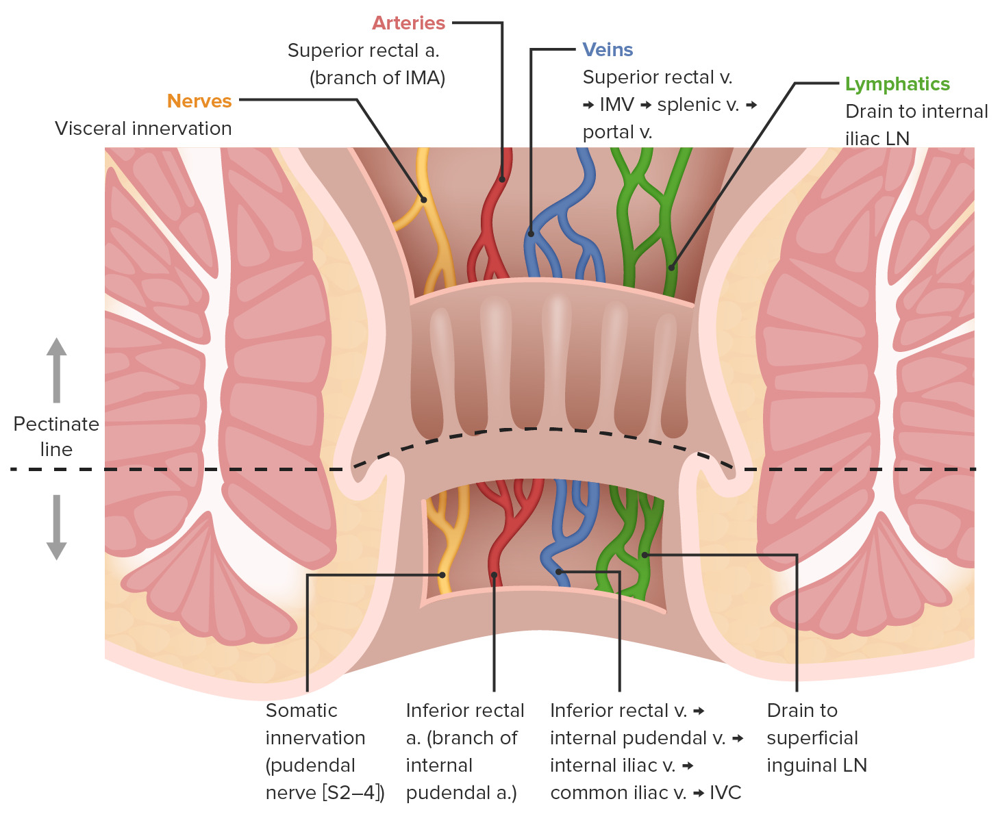
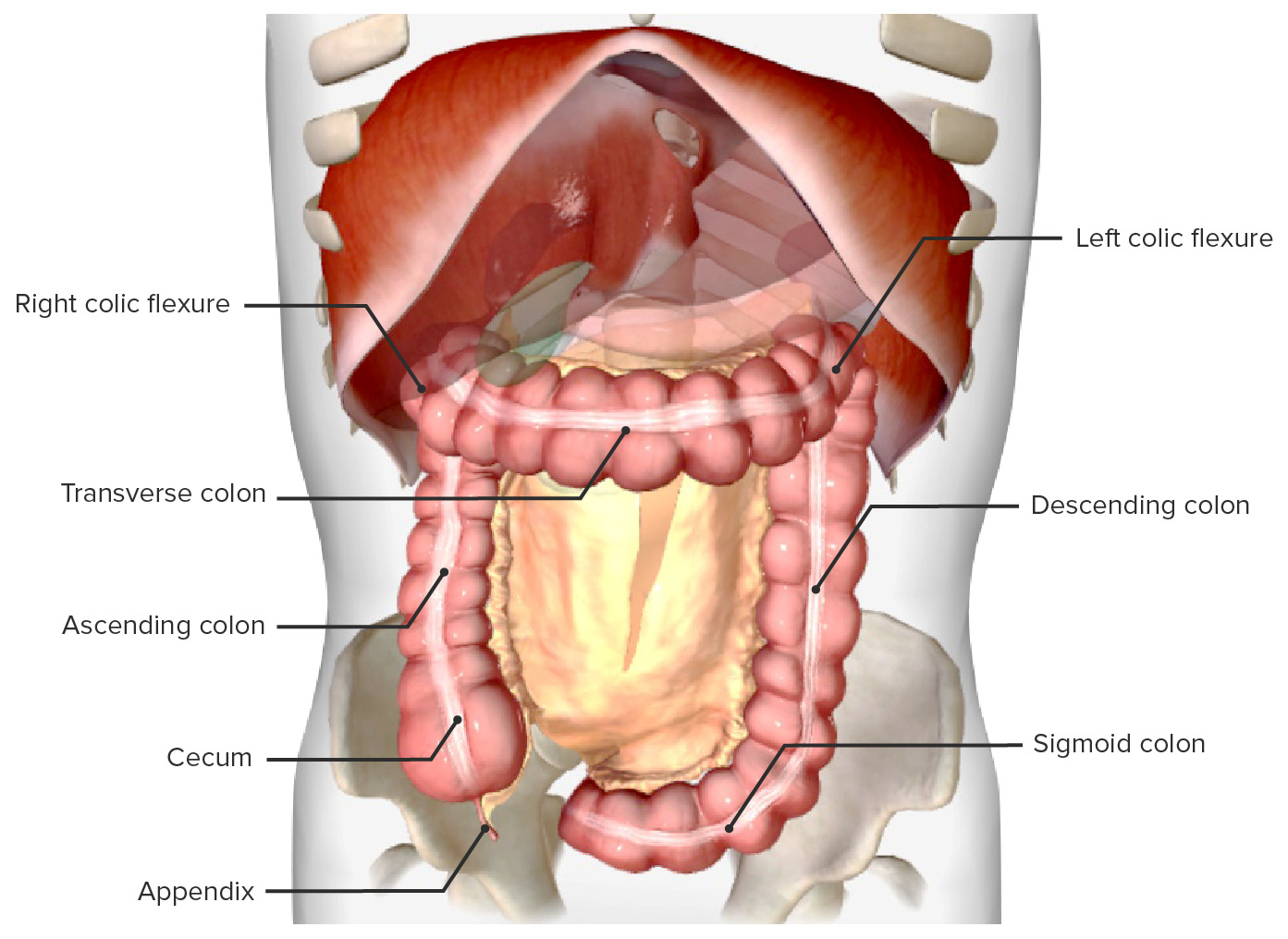
00:01 Let me now describe the histology of the large intestine. It's a fairly simple organ to describe histologically because its functions are merely to store the unwanted and undigested products as feces and also to absorb water. On the left hand section, you can see a low magnification section through the colon or large intestine. Identify the lumen and the folded mucosa. See if you can make out the submucosa, and then finally, the muscularis externa. The muscularis externa has a specialized longitudinal band called the teniae coli. 00:48 They're three bands running along the length of the colon. And when the colon is empty or almost empty, components can fold up, form circulations because of the contraction of these teniae coli. There are no villi in the large intestine, in the colon. The folds you see are submucosal folds, and the mucosa only consists of invaginations of the epithelial surface into glands. In the middle section, you can see an example of this. They're straight tubular glands. They're quite long. They embed deep into the lamina propria so that the cells there that are absorbing water, which is their function, are away from the hostile environment in the lumen. These glands are often referred to as the Crypts of Lieberkuhn. Besides having cells there, enterocytes there that absorb the water back into the body, there's a lot more goblet cells appearing now. 02:04 You see those nicely stained in that special section on the right hand side. Notice how many goblet cells there are. The epithelium is dominated in parts by these goblet cells. 02:16 They are going to lubricate the feces as they pass out of the body or help lubricate the feces. So really, the large intestine is a simple organ, stores unwanted products, undigested products, and absorbs water back into the system, and the mucus of the goblet cells helps to lubricate the feces. Finally, let's have a look at the rectum
About the Lecture
The lecture Large Intestine: Histology by Geoffrey Meyer, PhD is from the course Gastrointestinal Histology.
Included Quiz Questions
What are the three longitudinal ribbons of smooth muscle on the outside of the large intestine?
- Taenia coli
- Linea alba
- Linea semilunaris
- Rugae
- Muscularis mucosae
Author of lecture Large Intestine: Histology

Geoffrey Meyer, PhD
Customer reviews
5,0 of 5 stars
| 5 Stars |
|
5 |
| 4 Stars |
|
0 |
| 3 Stars |
|
0 |
| 2 Stars |
|
0 |
| 1 Star |
|
0 |
