Playlist
Show Playlist
Hide Playlist
Aortic Arches and Large Arteries: Introduction

-
Slides 06-32 Aortic Arches and Large Arteries.pdf
-
Reference List Embryology.pdf
-
Download Lecture Overview
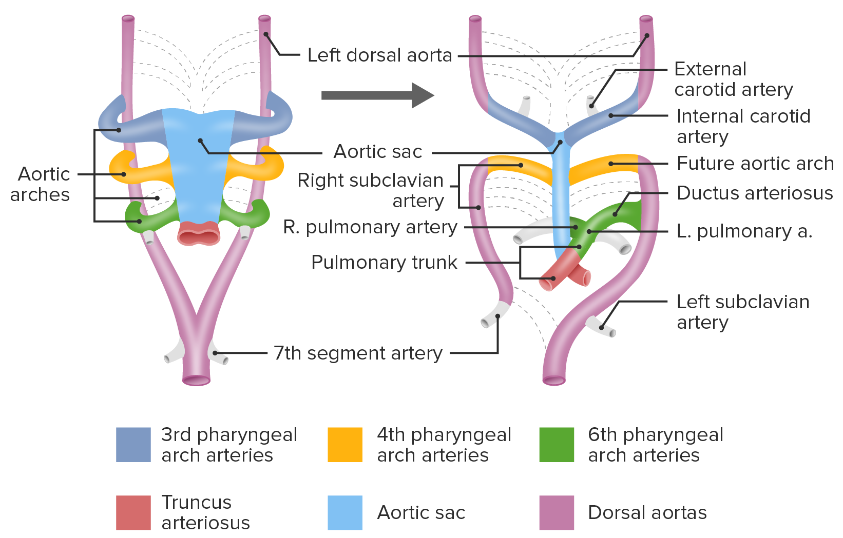
00:01 Hello and welcome to the first talk we're going to have on the development of the vascular system. 00:06 We're gonna start by discussing the aortic arches and very large arteries that are gonna be leaving the heart. 00:11 So it's hard to think about the time when we didn't have blood and blood vessels in our body but in early, early development we have our cells fed by diffusion from fluid filled chambers. 00:22 But as we get larger and larger we eventually need a circulatory system that allows us to get nutrients to and from the cells as our body gets larger and larger. 00:31 So formation of blood, blood vessels and the heart all have to work in close coordination so that when one is present, the others are present and the heart can pump and actually have something to pump with and through. 00:46 You're no good to have a heart if you don't have vessels and vice versa. 00:49 So at the very earliest time we're gonna start seeing vessels at around about day 17. 00:55 Little things called blood islands are starting to form inside the yolk sac. 01:01 Now, proper term would be angiogenic cell clusters, clusters of cells that are going to make blood. 01:06 And within day 18, we're gonna start seeing the same sort of things appearing in the mesoderm, both in the embryo and in the connecting stalk leading to the eventual placenta. 01:16 Now inside these angiogenic cell clusters, we have mesoderm that's starting to hollow out and create vessels and within them are little blebs of cells and these are the hematopoietic cell clusters that are gonna start generating the fetal blood supply, but not fetal blood supply, embryologic blood supply which then becomes the fetal blood supply. 01:38 Early on, this is where blood comes from but it's eventually gonna switch to the liver, spleen, thymus and bone marrow as those organs become available sites for hematopoiesis. 01:48 So let's talk about the vessels that leave the heart and we'll return and talk about the veins in the subsequent lecture. 01:56 The vitelline artery is the artery that supplies the yolk sac and while that's not gonna sound like a big deal right now, it's a very big deal in the early embryo and it's tied to the development of our gastrointestinal system. 02:09 The umbilical arteries are even more important because they're taking poorly oxygenated blood from the fetus to the placenta where it's gonna be oxygenated and then return through the umbilical veins. 02:23 More on those in just a little while. 02:25 And last but not least, we've got arteries that are distributing all over the body. 02:29 So the dorsal aorta on the right and left fuse into a single dorsal aorta but midway down the developing embryo and there are going to be little segmental branches that are leaving it all along its length supplying those tissues. 02:44 So we're gonna start by discussing how the heart pumps blood into the aortic sac and then distributes it to the dorsal aorta through aortic arches. 02:54 Now there are gonna be 1, 2, 3, 4, 6 aortic arches that develop in humans. 03:00 The 5th arch does not tend to form because humans do not have that 5th set of gill arches that some other animals do. 03:08 This is tied to the development of the pharyngeal arches and we have a separate lecture on that that you can check out if that sounds at all mysterious or unclear. 03:16 So I want you to note that the heart will pump blood in the aortic sac, the blood will then travel posteriorly through the aortic arches to reach the right and left dorsal aorta. 03:26 At that point, blood is gonna travel through intersegmental arteries to reach the various portions of the body that are developing like the neck, the torso, the limbs and so on and then a little further down it'll fuse to a single dorsal aorta. 03:41 One very important relationship right now is that the gut tube is forming posterior to the heart but it's gonna be travelling anterior to the dorsal aorta. 03:51 That's gonna be important when we discuss vascular slings and how malformations of the blood supply can occur. 03:58 Now if we take a look at this structure, the early aortic arches to my mind looked a bit like one of those facehugger aliens from the Aliens movies. 04:08 They are wrapping around to get to the dorsal aorta from the aortic sac. 04:12 The first and second arches are gonna tend to disappear very quickly and they don't contribute a great deal to the mature vasculature. 04:21 The 1st arch will remain as a bit of the maxillary artery and the 2nd arch will remain as a tiny little bit of the stapedial artery, the artery that supplies the smallest of the inner, pardon me'the middle ear bones, the stapedius. 04:34 The 3rd, 4th and 6th pharyngeal arteries are the ones that really make a big difference. 04:39 I want you to know that the 3rd arch, 4th arch and 6th arch all attach to the dorsal aorta, however, the 3rd and 4th arch are going to have their connection to the dorsal aorta separate. 04:52 So if I back up just a moment, you can see here that the dorsal aorta are continuous with all the arches feeding into each of them but as the bone proceeds the connection between the 3rd arch and 4th arch is going to disappear and the dorsal aorta will detach. 05:10 The 3rd arch is going to become the internal/external, but primarily the common carotid artery, so internal/external carotid arteries are branching off of it but the 3rd arch is gonna make the common carotid artery leaving towards the head. 05:26 The 4th arch is connected to a more posterior and inferior portion of the dorsal aorta, so it's going to arch out and head down the body. 05:35 And then the 6th arch does something very interesting. 05:38 It's going to disappear on the right, but retain it's connection to the dorsal aorta on the left. 05:44 So follow the 3rd arch going to the head. 05:48 4th arch and 6th arch stand a little lower down on the body and as we go a little further along, the 4th arch is going to stay connected to the right side which will become the right subclavian artery and on the left side it'll retain its connection to the dorsal aorta and become part of the aortic arch. 06:06 Now, before we move a little further along to the 6th arch I'd like to take a moment and look at how this blood supply interacts with the gut tube. 06:14 Remember that it's wrapping around the gut tube from anterior to posterior to reach the dorsal aorta. 06:20 As this is happening, the lungs are starting to develop off of the gut tube and the lung buds are gonna grow forward from the foregut and as that happens the blood supply associated with the developing lungs is going to be coming from the 6th aortic arch. 06:36 So the 6th aortic arch is going to give off the eventual pulmonary arteries. 06:41 So at this point on the picture on the right side of the screen, we can see that the pulmonary arteries are connecting to the 6th arch and the 6th arch is connecting to the dorsal aorta. 06:52 Eventually, we're gonna leave both pulmonary arteries uninterrupted but we're going to detach the right 6th arch from the dorsal aorta, whereas on the left it retains its connection to the dorsal aorta and becomes a structure called the ductus arteriosus.
About the Lecture
The lecture Aortic Arches and Large Arteries: Introduction by Peter Ward, PhD is from the course Development of Thoracic Region and Vasculature.
Included Quiz Questions
Which vessel supplies the yolk sac?
- Vitelline artery
- Umbilical artery
- Dorsal aorta
- Ventral aorta
- Aortic arch
Which pharyngeal arch fails to develop in humans?
- 5ᵗʰ arch
- 2ⁿᵈ arch
- 3ⁿᵈ arch
- 4ᵗʰ arch
- 6ᵗʰ arch
From which pharyngeal arch is the stapedial artery derived?
- 2ⁿᵈ arch
- 1ˢᵗ arch
- 3ʳᵈ arch
- 4ᵗʰ arch
- 6ᵗʰ arch
From which pharyngeal arch is the common carotid artery is derived?
- 3ʳᵈ arch
- 1ˢᵗ arch
- 2ʳᵈ arch
- 4ᵗʰ arch
- 6ᵗʰ arch
From which aortic arch are the pulmonary arteries derived?
- 6ᵗʰ arch
- 1ˢᵗ arch
- 2ⁿᵈ arch
- 3ʳᵈ arch
- 4ᵗʰ arch
Which vessel does the left 6ᵗʰ aortic arch give rise to?
- Ductus arteriosus
- Aortic arch
- Subclavian artery
- Stapedial artery
- Internal carotid artery
Author of lecture Aortic Arches and Large Arteries: Introduction

Peter Ward, PhD
Customer reviews
5,0 of 5 stars
| 5 Stars |
|
5 |
| 4 Stars |
|
0 |
| 3 Stars |
|
0 |
| 2 Stars |
|
0 |
| 1 Star |
|
0 |
