Playlist
Show Playlist
Hide Playlist
Pulmonary Embolism (PE): Imaging Studies

-
Emergency Medicine Bord Pulmonary Embolus.pdf
-
Download Lecture Overview
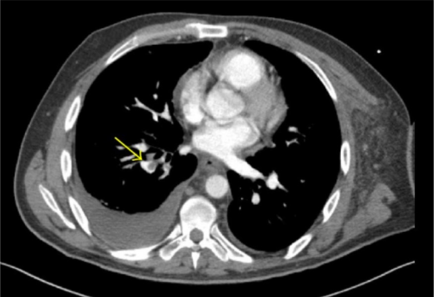
00:01 So focusing on these imaging studies, CT Angiography is a CT scan with IV contrast which looks at the blood vessels. 00:08 On this CT scan caught here, those areas of gray in a large pulmonary vessels are areas where there's blood clot present. 00:16 You know, I mentioned there are few criteria that you need in order to be able to get a CT Angiography. 00:21 You need to actually have a good IV in your antecubital fossa, and that helps time the IV dye. 00:27 You also need to not have renal failure, and you need to be able to lay flat for the CT scan. 00:33 One of the advantages of CT angiography is generally CT scan machines are located very close to the emergency department. 00:40 So it's easier to send someone who's a little bit more sick there. 00:43 Sending someone to nuclear medicine for a VQ scan oftentimes the nuclear medicine departments are further from the emergency department. 00:50 So you wanna make sure your patient is pretty stable before you send them there. 00:53 And it's a nuclear medicine study which looks for a mismatch of ventilation and perfusion. 00:58 Now, if there's a mismatch of ventilation and perfusion, then that patient potentially has a pulmonary embolus. 01:06 Now, when getting a VQ scan, it's important that the patient doesn't have any underlying pulmonary disease, because you can imagine if they have underlying pulmonary disease or a pneumonia that already might make interpretation of your VQ scan a little bit challenging. 01:19 The other thing to think about with the VQ scan is it comes back with sometimes nonspecific information, so VQ scans can either be normal, or they can read as low prob-- moderate probability, or high probability for PE. 01:33 So they don't necessarily always give you a slam-dunk answer. 01:37 So you're gonna have to interpret your results of your VQ scan in conjunction with the patient and their risks.
About the Lecture
The lecture Pulmonary Embolism (PE): Imaging Studies by Sharon Bord, MD is from the course Respiratory Emergencies.
Included Quiz Questions
Which of the following statements about CT angiography is INCORRECT?
- IV placement is not significant in a CT angiogram study.
- CT angiography uses IV contrast to look at the blood vessels.
- CT angiography requires the patient to lay flat during the CT scan.
- Gray areas in CT angiography may represent areas with emboli.
- CT angiography cannot be used in patients with renal failure.
Which of the following statements about VQ scan is INCORRECT?
- VQ scans are specific for pulmonary embolism.
- VQ scan is a nuclear medicine study which looks for mismatch of ventilation and perfusion.
- A mismatch of ventilation and perfusion may signify the presence of a pulmonary embolus.
- It is important that the patient does not have any other underlying pulmonary disease when undergoing a VQ scan.
- VQ scan results should be interpreted in conjunction with the patient’s risk for PE.
Author of lecture Pulmonary Embolism (PE): Imaging Studies

Sharon Bord, MD
Customer reviews
2,0 of 5 stars
| 5 Stars |
|
0 |
| 4 Stars |
|
0 |
| 3 Stars |
|
0 |
| 2 Stars |
|
1 |
| 1 Star |
|
0 |
I dont like the presenters voice. other than that is is ok
