Playlist
Show Playlist
Hide Playlist
Glioma: Genetic Risk Assessment

-
Strowd CNS Tumors Glioma.pdf
-
Download Lecture Overview
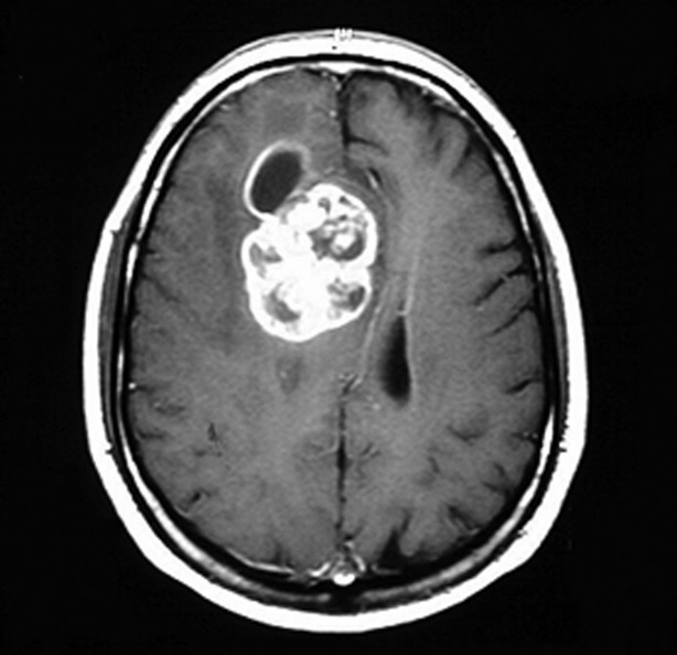
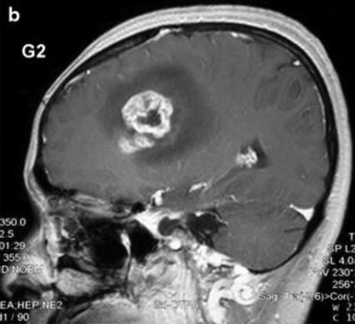
00:01 Histology is not the only way that we evaluate brain tumors. 00:04 Historically, histologic grading was the primary method for diagnosing tumors. 00:09 And in the last 10 to 15 years, we've learned that molecular assessments, looking at the tumors genes, gives us an important window into how tumors do, how patients will do, and how we should treat these tumors? I don't need you to understand all the details of this slide, but what we see here is that low-grade gliomas, things can go very differently depending on the type of low-grade glioma that it is, and the type of genes that we see in that tumor. 00:37 Here on the left side, we're looking at a Kaplan-Meier survival curve. 00:42 At the time zero point, the zero point of the X axis 100% of patients are alive. 00:48 And as we move forward in the graph, we see that patients succumb to their glioma and die. 00:54 We see that some patients do really well. 00:56 These IDH and TERT mutated tumors, a lot of patients are alive for a long time. 01:02 The same is true for triple positive tumors, not quite as much for IDH only, IDH mutant only tumors, not quite as much for the triple negative tumors, and not near as much for these TERT only tumors. 01:15 I don't need you to know all the details of the molecular features, except that genetic testing is important for determining how tumors will act, how aggressive they will be, how patients will do and what treatment we select? For high grade tumors, tumors that look like a glioblastoma under the microscope, molecular testing is also important, but it's not quite as helpful, as you see as for the low grade gliomas where the vast majority of patients, regardless of their molecular signature, will have a very aggressive course, regardless of the genetic events. 01:50 So we can put that all together and when we're evaluating a brain tumor, we start with the histologic appearance. 01:56 What did this tumor look like under the microscope? Did it look like an oligodendroglioma? A mixed oligoastrocytoma, which is a diagnosis that's falling out of favor? An astrocytoma or a glioblastoma? And we'll start our assessment of the tumor their. 02:13 Tumors that are histologically appear as an oligodendroglioma, or astrocytoma are termed diffuse gliomas. 02:19 And we start looking at key molecular events, genetic changes in the tumors, that will tell us, how that tumor will evolve and how that patient may do? The first gene we look at is something called IDH or Isocitrate dehydrogenase. 02:33 And it can either be mutant or wild type. 02:36 IDH mutant gliomas tend to respond better to treatment. 02:40 They tend to not grow as quickly. Patients tend to do better. 02:43 So we want to know about those. 02:45 IDH wild type tumors are less responsive to treatment. 02:48 And those patients sometimes the course can look very similar to a grade IV glioblastoma. 02:55 We can further subdivide these tumors based on other molecular events. 02:59 The IDH mutant tumors, those favorable risks likely to respond to treatment tumors, can acquire a loss of the 1 p arm. 03:07 the p arm of chromosome 1, or the q arm of chromosome2, which we call 1p19q codeletion, loss of both of those areas of the chromosome. 03:17 And those are oligodendrogliomas. 03:18 And they do exceptionally well with treatment and patients tend to do well with those tumors. 03:23 In contrast, IDH mutant tumors can acquire a mutation in the TP53 gene. 03:29 This is the most common gene to be mutated in all of cancer, and can be seen in gliomas. 03:34 And those tumors tend to take on the appearance of an astrocytoma and have an intermediate prognosis. 03:41 We said IDH wild type tumors can often be quite aggressive and not respond to treatment. 03:46 And are termed IDH wild type diffuse gliomas, and sometimes can act a very aggressive just like the grade IV glioblastoma. 03:54 Similarly, grade IV glioblastomas can be subdivided by their genes. 03:59 By that IDH gene as either IDH mutant gliomas or IDH wild type glioblastomas. 04:04 So you can see how our pathologist, and our tumor surgeons, and neurologist, start with the histology, add in the genetics, and come up with a final diagnosis of one of these five major types of gliomas.
About the Lecture
The lecture Glioma: Genetic Risk Assessment by Roy Strowd, MD is from the course CNS Tumors.
Included Quiz Questions
Which of the following genes is most commonly mutated in all cancer types and can be seen in astrocytomas?
- TP53
- 1p19q codeletion
- p16
- DPC
- WT1
[Select all that apply] Genetic testing is important in gliomas because it helps to determine ...
- ... how tumors will act (i.e., their behavior).
- ... whether the tumor has metastasized from another organ.
- ... the prognosis.
- ... the best treatment.
- ... the embryologic cell type.
Author of lecture Glioma: Genetic Risk Assessment

Roy Strowd, MD
Customer reviews
5,0 of 5 stars
| 5 Stars |
|
5 |
| 4 Stars |
|
0 |
| 3 Stars |
|
0 |
| 2 Stars |
|
0 |
| 1 Star |
|
0 |
