Playlist
Show Playlist
Hide Playlist
Formation of the Large Veins

-
Slides 06-33 Cardinal Veins and the Large Veins.pdf
-
Reference List Embryology.pdf
-
Download Lecture Overview
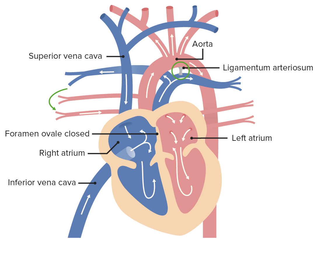
00:01 We're gonna continue our discussion of the development of the vascular system by looking at how the large veins form from a series of embryonic veins called the cardinal veins and the cardinal vein system. 00:11 Now, it would be sensible to think that the veins should simply parallel the arteries back into the body and that as the arteries develop out, the veins develop back in. 00:22 And that would make sense, but that's not what happens. 00:24 The venous system is gonna have a completely separate origin even though it drains the same areas and it's gonna have a very different pattern than the arteries that are supplying blood to the exact same area. 00:37 There are several large veins that are present early on in embryonic development. 00:42 The first is the vitelline vein. 00:43 It's gonna be draining blood from the yolk sac and even though the yolk sac isn't something we think of as being very important in human development, it is intimately tied with the formation of the GI system and the vitelline vein is gonna be tied to blood drainage from the GI system. 00:59 Now nearby, we have the umbilical vein. 01:01 Initially there are two umbilical veins bringing blood from the placenta back into the embryo. 01:07 But one is gonna dwindle, and we're left with a single umbilical vein. 01:12 And the most important thing about that blood is it's bringing oxygenated blood to the fetus. 01:18 The placenta's where gas exchange occurs while we are in the womb and this is the only way that the fetus is able to get fresh or at least partially fresh oxygen to perfuse the tissues and allow further development. 01:29 Last and certainly not least, we have a series of veins called the cardinal vein system. 01:35 And these cardinal veins all drain to what's called a common cardinal vein. 01:39 So there's gonna be an interior, posterior, and a variety of other cardinal veins that they all come together and enter the sinus venosus and initially, the umbilical and vitelline veins also drain to the cardinal vein and it's gonna be entering the sinus venosus of the heart. 01:56 The changes that occur in the cardinal veins, vitelline vein, and umbilical veins are gonna be what we spend the rest of this lecture discussing. 02:04 So initially, the umbilical and vitelline veins, as mentioned previously, drained the common cardinal veins to the sinus venosus, right horn and left horn, and then to the heart. 02:15 The vitelline veins are going to be surrounded by the liver. 02:19 The liver is growing off the gut tube and actually wraps around and surrounds the vitelline veins. 02:24 So these liver buds are gonna be wrapping around it and soon the vitelline veins will actually become the vascular spaces within the liver, but they retain their connection to the cardinal veins. 02:35 So the right and left vitelline veins are still draining blood into the common cardinal vein. 02:41 At the same time, the umbilical vein notices that the vitelline veins were there with the liver, and as the liver expands it also feeds into that system of vitelline veins. 02:53 And as it likes that pathway better it's gonna start losing its connection to the common cardinal vein. 03:00 So as we move to the next slide you'll see that the umbilical veins on either side flanking the gut tube are going to disappear between the liver and the common cardinal vein and their blood will thereafter simply travel to the liver. 03:13 So at this point, vitelline veins and the very proximal portion of the umbilical veins are both draining to the liver. 03:21 Common cardinal veins will be followed in a little more detail shortly. 03:24 Now, as development proceeds, the left umbilical vein starts to enlarge and the right umbilical vein starts to get a little bit smaller. 03:34 Notice that as we're following the changes that causes to the vitelline system. 03:38 The vitelline system is still within the liver and we're gonna wind up with a pathway between that enlarged left umbilical vein, enlarging, carrying it to the right side and the right vitelline veins. 03:51 We have a little bit of a diagonal pathway to the liver so that blood coming from the left umbilical vein doesn't have to filter its way through all the vascular space in the liver to get to the heart, it instead has a straight shot through the left umbilical vein and the structure that bypasses most of the liver called the ductus venosus, to the right vitelline vein, common cardinal vein, and then to the heart. 04:14 What's going to happen is once that enlargement occurs it just keeps going. 04:19 The left umbilical vein gets larger and larger; the right umbilical vein gets smaller and smaller; and the right vitelline vein, between the liver and the heart, is going to increase in size and the left vitelline vein in that same area will rescind and go away. 04:34 And we're left with something like this: The right umbilical vein is gone. 04:39 The left umbilical vein carries all the blood from the placenta, all that oxygen and blood into the liver to the ductus venosus, to the right hepatic vein, which is gonna be the remnant of that right vitelline vein into the inferior vena cava and then to the heart. 04:55 Nearby, the vitelline veins that were connected to the gut tube further down have formed some anastomotic connections between one another and are now forming what's called the portal veins or the hepatic portal veins. 05:09 These are the blood vessels that supply blood to the liver from the gut so the gut gets arterial blood, absorbs nutrients but our body wants the chance to detoxify and metabolize those structures. 05:24 So the hepatic portal system brings blood from the gut to the liver and that hepatic set of portal veins is coming from the vitelline veins, goes to the liver, goes into the vascular spaces, and then gets detoxified and then goes to the right, left, and middle hepatic veins to enter the inferior vena cava. 05:45 And at this point, we've more or less got the mature circulation of the liver and gut established with the exception of we're gonna not have blood flow in our umbilical veins during postnatal life.
About the Lecture
The lecture Formation of the Large Veins by Peter Ward, PhD is from the course Development of Thoracic Region and Vasculature.
Included Quiz Questions
Which embryonic vein carries deoxygenated blood from the yolk sac?
- Vitelline vein
- Umbilical vein
- Anterior cardinal vein
- Posterior cardinal vein
- Common cardinal vein
Which embryonic vein carries oxygenated blood from the placenta?
- Umbilical vein
- Vitelline vein
- Anterior cardinal vein
- Posterior cardinal vein
- Common cardinal vein
Which structure within the fetal liver allows blood from the left umbilical vein to reach the heart while largely bypassing the liver sinusoids?
- Ductus venosus
- Ductus arteriosus
- Vitelline vein
- Sinus venosus
- Left hepatic vein
From which structure do the liver sinusoids derive?
- Vitelline vein
- Umbilical vein
- Anterior cardinal vein
- Posterior cardinal vein
- Common cardinal vein
The hepatic portal vein is formed from what structure?
- Vitelline vein
- Umbilical vein
- Anterior cardinal vein
- Posterior cardinal vein
- Common cardinal vein
Author of lecture Formation of the Large Veins

Peter Ward, PhD
Customer reviews
5,0 of 5 stars
| 5 Stars |
|
5 |
| 4 Stars |
|
0 |
| 3 Stars |
|
0 |
| 2 Stars |
|
0 |
| 1 Star |
|
0 |
