Playlist
Show Playlist
Hide Playlist
Examination of the Head, Neck, and Ears

-
Reference List Physical Examination.pdf
-
Download Lecture Overview
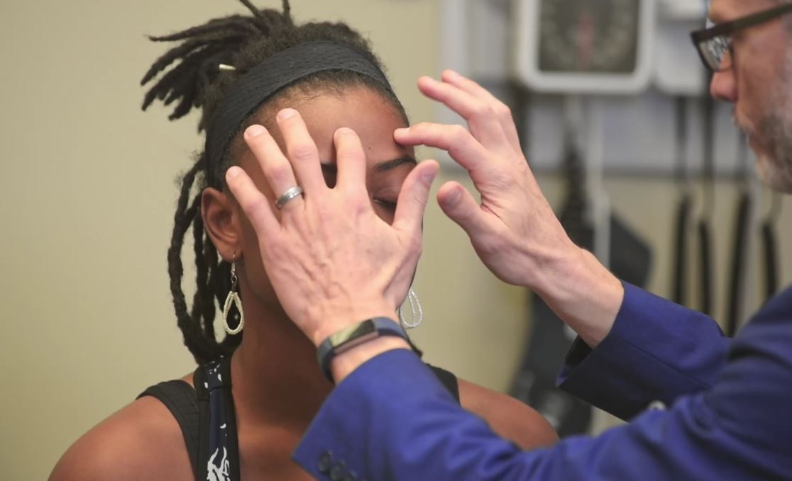
00:00 We're next going to move on with the head and neck exam. Some sections of this lecture are covered in more detail elsewhere for example the cranial nerves, the lymph node exam. 00:11 We're also going to be doing fundoscopy in a later lecture as well. So, there will be a little bit of redundancy later on in this course. And of course redundancy is very useful when it comes to learning stuff so that's okay. With that, we'll take a look first at gross inspection. 00:25 Oftentimes, you can detect some congenital abnormalities like Down syndrome, etc. simply by doing a quick inspection of the head with particular abnormalities in the nasolabial folds, in the lateral canthi of the eyes, the spacing of the ears and the eyes, etc. but we're just focusing on looking for any acute abnormalities at this time. Patients with parotitis, you'll see a little bit of this chipmunk kind of description they describe it with these bulging cheeks out to the sides just anterior to the ears and bilateral parotitis may be present in patients with mumps or alcohol use disorder due to malnutrition, a variety of different causes and you can look for that now. Otherwise, we can move on with percussing and palpating the sinuses. I'll just add that when you look at the eyes, there are also going to be a variety of manifestations of hyperthyroidism. Patients with proptosis, which just means that the eyelid is failing to drop over the iris. There's a little bit of the sclera that you're catching above the pupils, you could very quickly detect that by looking at the patient. And that's due to the increased sympathetic tone that accompanies hyperthyroidism. So I'm not seeing any of that, you can very clearly see that there's no sclera above her iris, her eyelids are perfectly resting where they're supposed to. So now I'm going to just percuss her frontal sinus and maxillary sinus, looking for any tenderness in those areas. Now we can move on to assess the ears. We'll take a look for external acoustic meatus issues, i.e. otitis externa versus otitis media. So for the assessment of otitis externa, it's going to start externally first. And so, just tugging on the auricle of the ear can sometimes elicit pain in a person who has otitis externa. And you can also palpate the tragus here and simply palpating that area may also cause a patient to have discomfort. Also, just in terms of looking for signs of infection, we would perform the lymph node exam. And in a later course we'll talk about that comprehensive lymph node exam, but you may just at least in this case want to palpate the preauricular and posterior auricular areas when you're thinking about an ear infection. Alright, so let's proceed with otoscopy to take a good look at the external acoustic meatus. Alright, you could turn towards me again please, Shayla. So what we're going to do now is just quickly orient you to your otoscope. It's a fairly straightforward device unlike the ophthalmoscope, which is a bit more complicated. The otoscope essentially usually has a green line here, which indicates that your practitioner who's using this device has 20/20 vision if they can line it up with the green line. Otherwise, if you're nearsighted or farsighted, you may have to dial it up or down and you'll know that when you start going into the ear or looking for the tympanic membrane. 03:16 Otherwise, you're simply dialing this up or down based on how much light you want to use in your otoscope. But in this case, you want to maximally use your light to perfectly visualize your tympanic membranes. So I hold my otoscope kind of like an upside down pencil and that allows me to have maximal control and I could also sort of steady my hand on the back of her head, and my other hand is going to pull up on her helix. By pulling up on the helix of the ear, you're raising up the peripheral part of the external acoustic meatus which allows for better visualization of the tympanic membrane since oftentimes I find the external acoustic meatus kind of drips down or kind of obscure the view. So, we'll pull up on the auricle here, I'm going in with my otoscope. And first I'm attending to the ear canal itself. And in this case, it is nice and pink, I can see hair cells. There's no wax in the ear. And I don't see any signs of erythema. I don't see any denudation of the skin. Oftentimes patients who are routinely using Q-tips are actually causing damage to the ear canal itself. So in this case, I see healthy ear canal and so I can move on and take a look at the tympanic membrane itself. Pulling up on the helix again. Now looking at her tympanic membrane, I see that it is nice and gray and translucent. I can see the light reflex which is reflected inferiorly and anteriorly, which is appropriate. I can see the ossicles, specifically the malleus up at the top. There's no evidence of erythema, there's no perforations on the tympanic membrane. There's no evidence of any vesicles or bubbles on the other side of the ear, and it's not bulging out towards me. So this tells me that this is a healthy tympanic membrane and a healthy middle ear.
About the Lecture
The lecture Examination of the Head, Neck, and Ears by Stephen Holt, MD, MS is from the course Examination of the Head and Neck Region.
Included Quiz Questions
A patient with bulging cheeks often described as “chipmunk” facies likely has which of the following diseases?
- Mumps
- German measles
- Adenovirus
- Roseola infantum
- Measles
On physical examination, a patient's sclerae can clearly be seen above their irides. What condition does this patient most likely have?
- Hyperthyroidism
- Hypothyroidism
- Cirrhosis
- Addison's disease
- Wilson disease
When using an otoscope, what does the green line represent?
- The appropriate setting for a user with 20/20 vision
- Maximum intensity light
- Minimum intensity light
- The otoscope has recently been calibrated.
- The otoscope is fully charged.
What physical exam finding is most suggestive of otitis externa?
- Pain when pulling on the ear
- Bulging tympanic membrane
- Absence of the light reflex
- Erythema of the tympanic membrane
- Nonerythematous external acoustic meatus
Author of lecture Examination of the Head, Neck, and Ears

Stephen Holt, MD, MS
Customer reviews
3,8 of 5 stars
| 5 Stars |
|
3 |
| 4 Stars |
|
0 |
| 3 Stars |
|
1 |
| 2 Stars |
|
0 |
| 1 Star |
|
1 |
Incredible lecture series and clear explanations, a total must for physicians regardless of the level of knowledge!
Thank you for the comprehensible content. Very useful. Totally worth it.
3 customer reviews without text
3 user review without text
