Playlist
Show Playlist
Hide Playlist
Examination of the Breast

-
Reference List Physical Examination.pdf
-
Download Lecture Overview
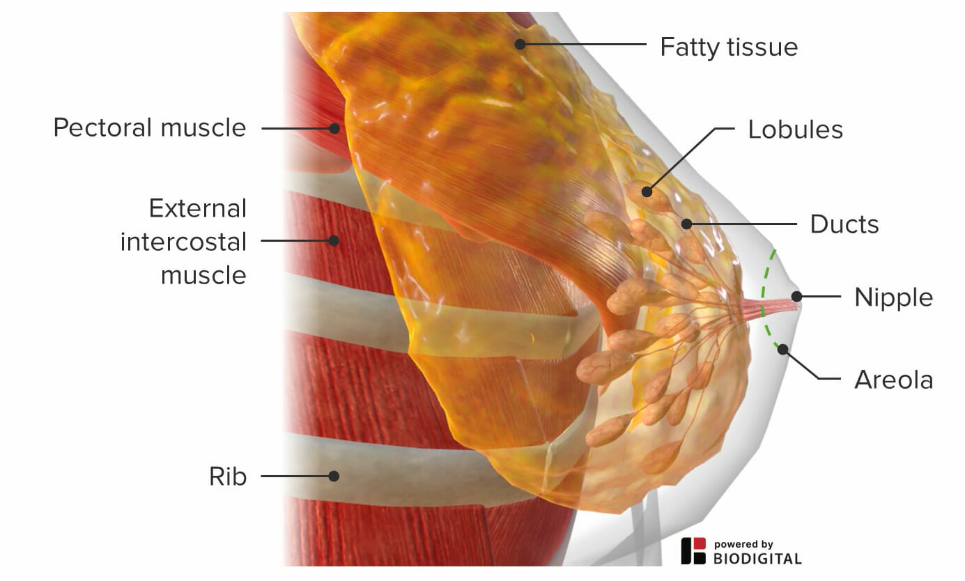
00:00 So now we're going to move on with the breast exam. 00:03 Now, it's important to recognize that the breast exam, like the genital exam, is a very sensitive part of the exam and it's important to walk through the entirety of the exam with your patient in advance to make sure they know what to expect. 00:14 It's also important to have a chaperone in the room with you. 00:17 The last thing that I'll just mention is that sometimes when you're palpating the breast, if you have, if the patient has fibroadenomas or other benign lumps, they can be more tender or painful during menses. 00:27 So if it's possible to time the exam after menses, that can be a bit more comfortable for the patient. 00:35 All right, with that, we'll move on to inspection of the breasts. 00:37 So Shayla, if you're okay with it, if you wouldn't mind just lowering your gown. 00:43 So on inspection, you're mostly just looking for symmetry, though, acknowledging that oftentimes one breast may be slightly larger than another and that's not unusual. 00:52 In order to get a full view of the contours of the breast, I'll just ask you to lift up your arms. 00:57 That allows us to see each quadrant of the breast. 01:02 And we'll talk about the quadrants a bit more detail in a moment. 01:04 We're also looking for any evidence of nipple retraction or an dimpling of the skin, which may be indicative of an underlying breast cancer, which oftentimes a breast cancer close to the surface of the skin will pull the skin in. 01:17 And there's also the peau d'orange type deformation that you can also see with certain types of breast cancers. 01:23 I'm also looking for just any skin changes at all. 01:26 Patients with mastitis in the setting of inflammation during breastfeeding, and you could also see that at this point as well. 01:32 You can lower your arms now please, Shayla. 01:35 And with that, we will go ahead and move on with palpation of the breasts. 01:40 So I'll take this opportunity just before I move on to the remainder of the breast exam to highlight the idea that we don't need to wear gloves when you're performing most of the physical exam, unless you're dealing with an open wound or obviously examining the genitalia, there's no reason to put gloves on. 01:56 All they're going to do is reduce the sensitivity of your finger pads, the most sensitive part of your fingers, and obscure things like in this case, we're looking for little lumps and bumps in the breasts. 02:06 So don't throw gloves on just because you're examining another human being, instead, just wash your hands. 02:16 All right, so with that, may I move one side of the sheet aside? I find it's useful to just examine one breast at a time to maximize the comfort and modesty of your patient. 02:28 And what I'll do is bring your arm a little bit to the side, just like that When we're examining the breast, typically, we think of the breast as divided into four quadrants. 02:36 There's, of course, an upper inner quadrant, upper outer, and then you've got a lower outer and lower inner quadrant as well. 02:43 And then this is the tail of Spence, which is glandular tissue that is heading up towards the axilla. 02:49 The typical approach that we use for examining the breast, there's two different types. 02:53 There's kind of a going in a circle approach and then an approach that appears to be somewhat more evidence-based is called the "lawnmower approach" in so far as you just go up and down with vertical lines, one after the other, and you're perhaps more likely to catch every single part of the breast if you follow that methodical approach. 03:13 So I'm going to lay hands on, you know, if it's okay and we're going to just start on the medial aspect of the breast. 03:18 I'm going to displace your breast a little bit up here as I do that. 03:24 And I do this kind of little superficial circle and then deeper circle approach. 03:36 And I'm going to come back the other way. 03:41 It's not unusual, particularly around the time of menses, to find benign lumps, fiberoadenomas which tend to be a bit more tender and a bit more enlarged during menses . 03:56 And the characteristic features of lumps that you're looking for and you want to characterize are, is the lump round? Lesions that are fairly symmetrical and round are more likely to be benign. 04:10 Is it mobile? A lesion that is fixed to the skin or to the anterior chest wall is more likely to be cancerous. 04:19 Tenderness, as I alluded to, a tender lesion is less likely to be cancers as well, whereas a firm fixed asymmetric lesion is one that we're more concerned about cancer. 04:47 Again, superficial and then deeper. 04:53 Just making sure I'm catching all of the breast. 05:22 Sometimes the border between the end of glandular tissue and the anterior chest wall can be subtle, so it's important to go beyond that border to make sure you don't miss anything. 05:34 And then I'm going to follow, like I said, the tail of Spence up here towards the axilla. 05:44 You're also going to want to perform a lymph node, exam in the axilla. 05:47 And the lymph node exam for the axilla is divided into four quadrants. 05:50 I like to think of it as a box with an anterior, posterior, lateral, medial wall. 05:54 I'm going to start here by just going underneath your armpit, if that's okay, Looking for bumps there. 05:59 I'm going to do the anterior wall where the pectoral muscles are, the chest wall, which is the medial side, then the posterior wall where your latissimus dorsi are. 06:09 Last you want to look at the nipple itself looking for any asymmetry around the nipple or any dimpling. 06:14 Just to mention here that male patients who have gynecomastia will actually have a similar increase in glandular tissue around the nipple as opposed to with obesity. 06:26 There tends to be just very soft adiposity in that area, whereas with gynecomastia, there really is a firmness, as if you're palpating glandular tissue. 06:35 And now I'll just going to squeeze at the base of the nipple to look for any expression of any discharge. 06:40 Patients who have breast cancer, that's glandular in type may actually, you'll express some blood or have some other secretions when you do so. 06:50 And there's no evidence of anything when I do that here on the breast. 06:55 So that completes the breast exam.
About the Lecture
The lecture Examination of the Breast by Stephen Holt, MD, MS is from the course Examination of the Breast and Lymph System.
Included Quiz Questions
Which feature of a breast lump is more concerning for a malignant than a benign process?
- A fixed mass that is not mobile
- Clearly delineated margins
- Symmetric findings in the other breast
- Slow growth
- Soft and rubbery consistency
What feature of breast tissue in a man favors benign tissue over the diagnosis of gynecomastia?
- Soft consistency of the adipose tissue
- Firm glandular tissue
- Most often found in adolescents and individuals > 50 years
- Related to hormone changes
- Tender breast tissue
What potential finding on breast/nipple exam is the most concerning for a malignant process?
- Bloody discharge from the nipple
- Axillary lymph nodes measuring < 1 cm
- Clear discharge from the nipple
- Inverted nipples
- Generalized breast tenderness a few days before menses
Author of lecture Examination of the Breast

Stephen Holt, MD, MS
Customer reviews
5,0 of 5 stars
| 5 Stars |
|
5 |
| 4 Stars |
|
0 |
| 3 Stars |
|
0 |
| 2 Stars |
|
0 |
| 1 Star |
|
0 |
