Playlist
Show Playlist
Hide Playlist
Esophageal Rings esp. Schatzki Ring

-
Slides GD Esophagus.pdf
-
Download Lecture Overview
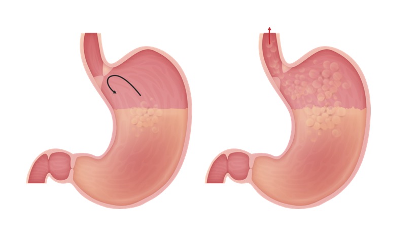
00:01 We move from cancer now into a ring. 00:05 What you want to keep in mind, diagnostic purposes, so that you get your questions correct, is keep your ring separate from your webs. 00:14 Keep your rings separate from your webs. 00:18 Begin our discussion with what’s known as a Schatzki ring. 00:21 A Schatzki ring. Your patient, who is he or she, greater than 40, so still relatively young but greater than 40. 00:30 Where would you find this ring in the esophagus? Would it be more proximal or would it be more distal? It would be distal. 00:37 Next, if there is a ring that is involved, then you want to think of this as being a fixed constriction of your esophagus in which the dysphagia would be to solids and liquids basically at the same time. 00:51 So therefore, the description of this clinically is called non-progressive dysphagia. 00:57 When are you thinking… So henceforth, whenever you hear the word dysphagia, do you understand now that you’re looking for descriptions around that dysphagia. 01:07 Is it progressive? Is it non-progressive? Is it at the same time? So on and so forth. 01:12 This is going to give you the answer. 01:15 Exacerbation with what’s known as hurried eating and possibly development of later on, reflux. 01:25 The squamous epithelium on the upper surface, is a columnar on the lower surface. 01:30 Where are we when we talk about Schatzki ring? You’re at the lower esophagus. 01:36 When you’re at the lower esophagus, then you’re that much closer to the stomach. 01:40 You should know the general histology of your esophagus. 01:43 So think of it as being your squamous. 01:45 Now keratinized, stratified, so on and so forth but squamous is what you’re paying attention to, is that clear? Whereas if you’re moving into the stomach, then you have the columnar type. 01:56 Remember that because you’re down in the lower esophagus, there’s every possibility that over a long period of time that reflux clinically is part of this syndrome. 02:06 So if reflux is also taking place simultaneously with this ring, do you understand that now because the ring literally behaves kind of like a panel, in which the underside of it is being literally hit by reflux. 02:22 But the underside which used to be squamous, is undergoing a metaplastic process. 02:28 Therefore the underside of the ring will be columnar in nature. 02:33 Whereas the upper portion of the ring is going to be more of your normal esophageal histology, will be squamous. 02:42 Use common sense. 02:43 Understand that you’re by the lower esophageal sphincter, with the accompanying reflux, the underside would be more so of the columnar nature, whereas the upper side will be more of your squamous. 02:57 Found 6% to 14% of your population, treatment, here once again, numerous ways of trying to dilate your esophagus. 03:06 Repeated due to recurrence. 03:08 That’s the only thing with rings because you… yes there’s every possibility of going on to cancer, dysplasia, anytime there’s irritation. 03:15 Anytime there’s irritation, may go on to dysplasia. 03:18 What kind of cancer? Do not worry about that. 03:22 What you’re paying attention to is diagnose the ring first and once you do so, you do everything in your power to make sure that you dilate this esophagus. 03:30 So you are relieving the symptoms of the patient. 03:34 For Pete’s sakes, let the patient eat. 03:39 Dilatation, but you need to repeat it because of recurrence. 03:43 Let’s take a look at the ring here. 03:45 The ring, so this is the second time in which we’re looking at a, endoscopic examination. 03:51 The first time I showed you endoscopic examination, I began a discussion of strictures. 03:56 You’ll notice here on your right, you do an upper endoscopy, you’re scoping your patient. 04:02 Okay? And you have a little camera in which you’re looking at the esophagus. 04:06 You look at the lining of the esophagus, there is no stricture here. 04:10 This is no varices. 04:11 This is no infection. 04:13 This is not Barrett’s tongue. 04:15 This is actually pretty much on top of the ring. 04:19 Normal histology. 04:20 Do you see how fleshy, and beau-… I mean, I think this is beautiful. 04:24 I think is… For the most part this is normal. now where are you? Now take a look at the left picture with the imaging, and you have your barium swallow. 04:33 With that barium swallow on esophagram, you’ll notice that the very bottom, so you’re moving down to the bottom of the tube, please. 04:41 And you see an area there which is literally become compromised. 04:45 So you’re by the lower esophageal sphincter. 04:48 In other words, you’re in the distal esophagus. 04:50 Histologically, may I ask you a question? On top of the ring what kind of histology would this be? More normal type, so there… more normal of the esophageal type, so it’ll be squamous. 05:02 Underside if there’s reflux associated with it, then it would be more of your columnar cells. 05:08 Imagine here once again, the lumen has become compromised and so therefore making it difficult for the patient to swallow solids and liquids. 05:17 Rings. 05:20 Schatzki ring is what we talked about. 05:23 A fixed stricture, or I have to be careful, I wouldn’t call it a stricture, but a fixed obstruction is a better term for this. 05:30 A stricture, I showed you earlier was due to, let’s say injury taking place in esophagus, in the lower esophagus.
About the Lecture
The lecture Esophageal Rings esp. Schatzki Ring by Carlo Raj, MD is from the course Esophageal Disease: Basic Principles with Carlo Raj.
Included Quiz Questions
A 45-year-old patient presents to your office complaining of dysphagia. She states that she mostly has trouble with solids and that her dysphagia is intermittent. She says her condition is not getting worse but is continuing to frustrate her. Which of the following is the MOST likely etiology of her dysphagia?
- Esophageal ring
- Carcinoma of the esophagus
- Infection
- Chronic inflammation
- None of the above
What kind of epithelium would the histopathology of esophageal rings show under the microscope?
- Squamous cell epithelium on the upper surface and columnar epithelium on the lower surface
- Columnar epithelium
- Cuboidal epithelium
- Ciliated columnar epithelium
- Squamous cell epithelium
What is the MOST appropriate treatment for a Schatzki ring?
- Dilatation
- Smoking cessation
- Anti-inflammatory drugs
- Anti-cancer drugs
- None of the above
Author of lecture Esophageal Rings esp. Schatzki Ring

Carlo Raj, MD
Customer reviews
5,0 of 5 stars
| 5 Stars |
|
5 |
| 4 Stars |
|
0 |
| 3 Stars |
|
0 |
| 2 Stars |
|
0 |
| 1 Star |
|
0 |
