Playlist
Show Playlist
Hide Playlist
Erythrasma in Darker Skin
-
Slides Erythrasma in Darker Skin.pdf
-
Download Lecture Overview
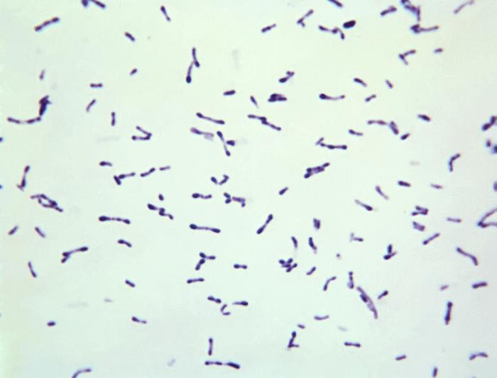
00:01 Welcome to our lecture on Erythrasma. 00:05 What is erythrasma? It's a superficial skin infection caused by Corynebacterium minutissimum. 00:14 It frequently occurs with a prevalence of approximately 4%. It's present globally, but particularly prevalent in subtropical and tropical regions. 00:27 It's less common in children, and the incidence is higher in black patients. Some of the risk factors include older patients, patients with diabetes mellitus, and any condition that predisposes to skin occlusion and moisture. 00:45 Obesity is one of the risk factors of erythrasma. 00:48 And of course, hyperhidrosis, which is increased sweating. 00:53 Living in tropical climates increases the risk factor of developing erythrasma. 01:03 So Corynebacterium minutissimum is a gram positive bacillus that causes erythrasma. 01:10 It is part of the normal skin flora, but some factors, such as heat and humidity, induce the proliferation of the bacteria in certain individuals. They multiply within the stratum corneum, which is the upper layer of the skin, resulting in thickening and the distinctive clinical presentations on the skin. 01:31 Patients with erythrasma may present with the following clinical manifestations, and some of the predilection sites include the axillae, groin and toe webs. So what about the clinical manifestations? It's usually asymptomatic. 01:48 Patients don't complain of anything, but it may present with slightly scaly, brown, sharply demarcated plaques. 01:57 Physical examination. When you examine the patient, you will see scaling and maceration in the sites that I've just mentioned. If you use Wood's lamp, you may see coral red fluorescence, which confirms the diagnosis of erythrasma. 02:13 Hence erythrasma. Because the fluorescence is red on Wood's lamp. If one does a gram stain of a skin scraping, you see gram positive filaments and rods consistent with the detection of the bacteria. One of the differential diagnoses includes seborrheic dermatitis, where you see erythema and greasy scales on the scalp, eyebrows, and nasolabial folds. 02:41 Interdigital tinea pedis can also be easily confused with a erythrasma or interdigital erythrasma, so if you want to differentiate between the two. If you do a potassium hydroxide preparation, this can help with the differentiation. 02:58 The management of erythrasma includes, if it's localized, you use topical therapy with antibacterial agents as listed. 03:07 There's fusidic acid and macrolides. 03:10 And if it's extensive oral antibiotic therapy, for example, the macrolides and tetracyclines can be used.
About the Lecture
The lecture Erythrasma in Darker Skin by Ncoza Dlova is from the course Bacterial Skin Infections in Patients with Darker Skin.
Included Quiz Questions
What characteristic finding is seen when examining erythrasma lesions with a Wood's lamp?
- Coral red fluorescence
- Blue-green fluorescence
- Yellow fluorescence
- White reflectance
- No fluorescence but enhanced skin markings
Which microorganism is the causative agent of erythrasma?
- Corynebacterium minutissimum
- Staphylococcus aureus
- Malassezia furfur
- Trichophyton rubrum
- Pseudomonas aeruginosa
Customer reviews
5,0 of 5 stars
| 5 Stars |
|
5 |
| 4 Stars |
|
0 |
| 3 Stars |
|
0 |
| 2 Stars |
|
0 |
| 1 Star |
|
0 |


