Playlist
Show Playlist
Hide Playlist
Enamel, Dentin and Cementum

-
Slides Digestive system oral cavity.pdf
-
Reference List Histology.pdf
-
Download Lecture Overview
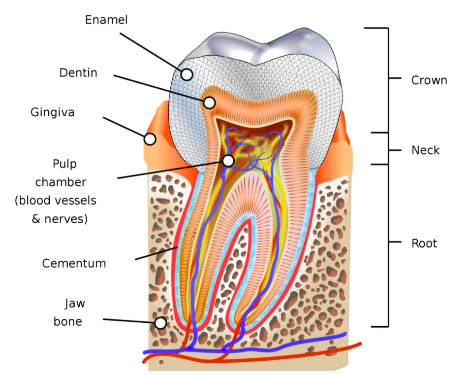
00:01 In this section, I want to just briefly explain the structure of enamel. It's a very hard structure. It's the hardest structure in the body. It consists of 98% crystals, calcium hydroxyapatite crystals, secreted during development from ameloblasts. And these ameloblasts sit on the surface of the junction between the dentin and the enamel with odontoblasts. And they sit like my hands are sitting close together here. 00:38 And during development, the odontoblasts secrete dentin and move towards the pulp cavity. The ameloblasts secrete enamel and they move to the exterior surface of the tooth during development. 00:54 And those ameloblasts lay down these enamel rods which can extend about anything from, they're really very long structures. The thickness of the crown of the tooth or the enamel is about two millimeters. So these enamel rods produced by the ameloblasts can be two millimeters in length. If you looked at them under very high power, they are key-shaped structure, and they all interlock making this structure a very very hard structure. It's the hardest structure in the body. Much harder than bone. And what the funny thing about it is, these ameloblasts are not connective tissue cells. Most of us realize that connective tissue cells lay down cartilage and bone, the hard connective tissues that we think about in the body. 01:51 Here, the hardest component, the hardest structure in the body enamel is actually produced by epithelial cells. Sometimes just underneath the cusps of these teeth, you'll find the rods are in different directions. It's called gnarled enamel. And that adds a bit of strength to the cusp area, but it also just represents the orientation of the enamel rods as they go to the circumferential sort of arrangement in shape of the cusps. Let's have a look at dentin in the center supporting enamel, and also cementum, which forms a very thin surface on the root of the tooth. Dentin is produced in a similar way to the enamel rods. As I showed you before, the odontoblasts secrete the dentin. 02:52 And as they secrete the dentin, they leave a process, a cell process behind, and that stays within these little tiny canaliculi called dentinal tubules, a bit like canaliculi in bone. So when you look at the thickness of dentin, you'll see these dentinal tubules that have the processes of the odontoblasts, which are located in the pulp cavity on the inner surface of the dentin that I'll show in a moment. They're processes of these odontoblasts, sometimes they're very branch at the surface. And they can take sensory information to nerve fibres connecting to them in the pulp cavity. On the right-hand image, you can see a section through the cementum, the very thick, almost bone-like structure coating the root of the tooth. And if you look carefully into this image, you can just make some sort of spidery type processes. They are just the cementocyte sitting in the cementum extending their processes out to get nutrition just like the osteocytes do in bone. Cementum is essentially bone. Dentin is harder than bone, but not as hard as enamel. It only contains about 70% of calcium hydroxyapatite crystals. Here, you can see the odontoblasts. 04:29 On the left-hand side, the tooth. The dark pink reddish area is the dentin. 04:36 The pale area again is the pulp cavity. You don't see the enamel. 04:39 In the pulp cavity, you can see the pulp cavity on the right-hand side section. The purply area is the dentin. 04:52 So, on the surface of the dentin, the inner surface against the location of the pulp cavity, are these odontoblasts. And if you look very carefully, you can see the processes of these odontoblasts extending all the way to the surface of the dentino-enamel junction. And these are within those dentinal tubules I explained earlier. If you look again very carefully, you can see a rather pale region of the dentin before you see that dark purply region. That's called predentin. 05:28 Those odontoblasts have just laid down that newly formed dentin, but it hasn't yet been mineralized. So it doesn't take out that purply stain you see in the more mature mineralized dentin. On the right-hand side again, just have a look at the pulp cavity. It contains enormous numbers of blood vessels, supporter cells, and neurofibers that carry sensations away from the tooth.
About the Lecture
The lecture Enamel, Dentin and Cementum by Geoffrey Meyer, PhD is from the course Gastrointestinal Histology.
Included Quiz Questions
Which of the following statements regarding the tooth enamel is correct?
- The tooth enamel is predominantly made of calcium hydroxyapatite produced by epithelial cells called ameloblasts.
- The tooth enamel is predominantly made of calcium hydroxyapatite produced by connective tissue cells called ameloblasts.
- The tooth enamel is predominantly made of calcium hydroxyapatite produced by epithelial cells called odontoblasts.
- The tooth enamel is predominantly made of calcium pyrophosphate produced by epithelial cells called ameloblasts.
- The tooth enamel is predominantly made of calcium pyrophosphate produced by connective tissue cells called odontoblasts.
Which of the following statements regarding the histological architecture of the tooth is CORRECT?
- The dentinal tubules are cellular processes of the odontoblast that extend into the dentinoenamel junction.
- The dentinal tubules are extracellular rod-shaped structures around the odontoblast that extend into the dentinoenamel junction.
- The odontoblasts are connective tissue cells located on the thin outer surface of the cementum.
- The cellular processes of the cementocytes carry sensory information to the nerve fibers in the pulp cavity.
- The dentinal tubules are cellular processes of the ameloblast that extend into the dentinoenamel junction.
Which structure in the tooth contains the blood capillaries and nerve fibers?
- Pulp cavity
- Enamel
- Dentin
- Predentin
- Cementum
Author of lecture Enamel, Dentin and Cementum

Geoffrey Meyer, PhD
Customer reviews
5,0 of 5 stars
| 5 Stars |
|
1 |
| 4 Stars |
|
0 |
| 3 Stars |
|
0 |
| 2 Stars |
|
0 |
| 1 Star |
|
0 |
It was very informative! I liked the explanation a lot.
