Playlist
Show Playlist
Hide Playlist
Ear: General Structure

-
Slides Vestibular system.pdf
-
Reference List Histology.pdf
-
Download Lecture Overview
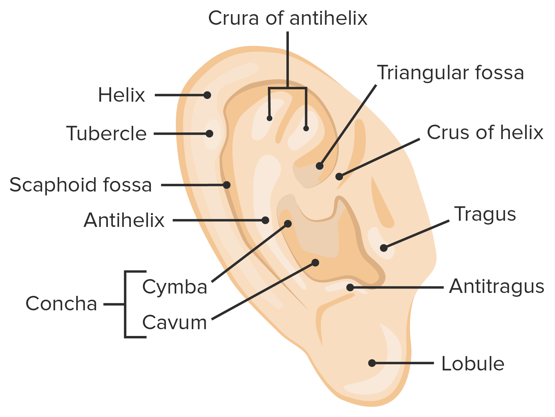
00:00 Let's, first of all, look at the general anatomical structure of the ear. On this diagram, there are various labels that do illustrate all the major components of the ear. Starting on the left-hand side is the pinna of the ear. That part of the ear is very flexible. 00:27 We can twist and bend it and it will spring back to its normal shape because it has got elastic cartilage in it. That helps to locate or amplify the sound but has very less function in humans than it does in other animals. The external auditory meatus is about two and a half centimeters long. It is bound on the most lateral part by the pinna of the ear. 00:58 And the very first third is supported by cartilage, and that cartilage blends with the elastic cartilage in the pinna of the ear. The skin, lining the external auditory meatus, has got special glands in it called ceruminous glands. Those ceruminous glands secrete a component that combines with apocrine secretions to produce ear wax. And that ear wax is used to try and trap any particular matter that happens to find its way into that auditory canal. And that ear wax is also suspended on the fine hairs in that canal again to increase the opportunity to trap that matter. On the medial aspect or the end part of that auditory tube is the tympanic membrane or the eardrum. It vibrates in response to sound waves. 02:04 And those vibrations are passed on to the bony ossicles, the malleus, incus, and stapes, illustrated in the diagram. And those bony ossicles will vibrate themselves, and those vibrations will then be passed to the oval window in the inner ear. The oval window and the round window play a very important role in the functioning of the inner ear. 02:34 You also have labelled there the auditory tube. It's a passage going from the middle ear cavity, all the way down to the pharynx. Normally, it's closed, except when we swallow or maybe when we yawn. It's a passageway or a potential passageway for pathogens passing out from the pharynx.
About the Lecture
The lecture Ear: General Structure by Geoffrey Meyer, PhD is from the course Sensory Histology.
Included Quiz Questions
Where is the tympanic membrane located?
- Junction of outer and middle ear
- Outer ear
- Middle ear
- Junction of middle and inner ear
- Inner ear
The auditory tube (Eustachian tube) connects which of the following structures?
- Middle ear and pharynx
- Outer ear and middle ear
- Inner ear and middle ear
- Inner ear and pharynx
- Outer ear and inner ear
Author of lecture Ear: General Structure

Geoffrey Meyer, PhD
Customer reviews
5,0 of 5 stars
| 5 Stars |
|
5 |
| 4 Stars |
|
0 |
| 3 Stars |
|
0 |
| 2 Stars |
|
0 |
| 1 Star |
|
0 |
