Playlist
Show Playlist
Hide Playlist
Overview of the Dural Venous Sinuses

-
Slides 20 DuralVenousSinuses BrainAndNervousSystem.pdf
-
Reference List Anatomy.pdf
-
Download Lecture Overview
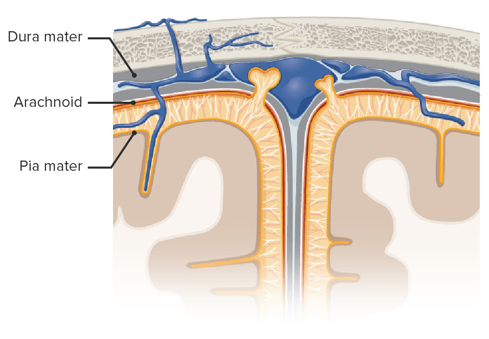
00:00 Welcome to this presentation on the dural venous sinuses. First, we need to understand some general considerations as they relate to this subject matter. Here is an example to begin of the superior sagittal sinus. 00:23 This is a very prominent sinus. This sinus as well as other sinuses are channels that form within the periosteal and meningeal layers of the dura mater. Where these sinuses is formed, there is a separation that allows the venous blood flow to circulate within the cranial cavity. These channels again, such as the superior sagittal sinus are lined by typical endothelium that you would see in any channel that is conveying or transmitting blood. The dural venous sinuses drain most structures of the cranial cavity. It’s important to understand that sinus venous blood drains primarily into the jugular veins. One of the features of the dural venous sinuses is the presence of arachnoid granulations. These are protrusions of the arachnoid mater through the dura mater into the venous sinuses. We have one labelled here in the illustration. 01:44 This is an arachnoid granulation. It’s projecting into the superior sagittal sinus. Then we see several others here as well. These are prominent in the lateral lacunae. We’ll see on the next slide where lateral lacunae are present. The reason we have arachnoid granulations is that they will effect the return of cerebrospinal fluid to the venous circulation. When we think about the dural venous sinuses, we also need to consider the arachnoid granulations. In this particular image, we can see several arachnoid granulations. 02:33 This area here is showing several prominent arachnoid granulations as well as this area here. These arachnoid granulations are located within the dura mater in areas called lateral lacunae. Lateral lacunae are areas where venous blood will be collected. The arachnoid granulations are returning cerebrospinal fluid to this venous blood. These lateral lacunae do communicate into this very large midline structure that’s referred to as the superior sagittal sinus. This slide introduces us to the various tributaries of the dural venous sinuses. 03:24 So, venous blood from elsewhere will drain into the sinuses. Of course, we also have the arachnoid granulations contributing as a tributary as well. The first tributary to keep in mind is that the bridging veins, these may be cerebral or cerebelli in origin. Here, we’re looking at a cerebral bridging vein. This one is emptying into the superior sagittal sinus. So that’s one type of tributary. A second type of tributary would be the presence of emissary veins. These are veins that run from the scalp, cross the skull itself, and can empty blood into dural venous sinus as we see here. Another type of tributary is the presence of diploic veins. 04:23 These are running within the skull as we see here and then can empty their venous blood into dural venous sinus. Meningeal veins are running within the dura mater as we see here. This meningeal vein is emptying in to this particular dural venous sinus. The last structure to point out on this image is that arachnoid granulations are also going to be tributaries. We have one arachnoid granulation here but they are a tributary for cerebrospinal fluid as it’s returning to the venous circulation.
About the Lecture
The lecture Overview of the Dural Venous Sinuses by Craig Canby, PhD is from the course Dural Venous Sinuses.
Included Quiz Questions
Which of the following statements about dural venous sinuses is NOT true?
- They are lined by columnar epithelium.
- Sinus venous blood primarily drains into the internal jugular vein.
- The presence of arachnoid granulations is one of their characteristic features.
- They arise from where the periosteal and meningeal layers of dura mater separate.
- They drain most structures of the cranial cavity.
Which statement describing arachnoid granulations is INCORRECT?
- They are protrusions of dura mater.
- They are protrusions of arachnoid mater.
- Lateral lacunae receive CSF from arachnoid granulations and drain into the superior sagittal sinus.
- They are located in the lateral lacunae.
- Arachnoid granulations effect return of CSF into the venous circulation.
Which of the following does NOT drain blood into the dural venous sinuses?
- Arachnoid granulations
- Emissary veins
- Cerebral bridging veins
- Meningeal veins
- Diploic veins
Author of lecture Overview of the Dural Venous Sinuses

Craig Canby, PhD
Customer reviews
2,3 of 5 stars
| 5 Stars |
|
1 |
| 4 Stars |
|
0 |
| 3 Stars |
|
0 |
| 2 Stars |
|
0 |
| 1 Star |
|
2 |
uhh uhh uhh uhh uhhh uhhh uhhh uhhh this is so disturbing
Excellent overview. Dr. Canby is one of my favorite lecturers for his clarity and straight-to-the-point lectures.
Unbearable. Even in 1.25x he speaks way too slowly. Boring tone of voice as well
