Playlist
Show Playlist
Hide Playlist
Development of the Teeth
-
Slides Head Neck Anatomy Teeth.pdf
-
Download Lecture Overview
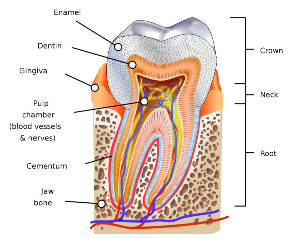
00:00 And so now that we understand the three regions of a tooth, and we understand the four types of tissues that constitute an adult tooth, now is the time to look at how a tooth develops. 00:17 There are six stages of tooth development, and I'll just rattle them off quickly here to begin, and then we'll revisit each one and layer in some details. 00:31 The first stage of tooth development is known as the "Bud stage". 00:36 The second stage is referred to as the "Cap stage". 00:41 Stage three is referred to as the "Bell stage". 00:47 Fourth stage is the "Secretory stage". 00:52 Fifth stage is Eruption of the developing tooth. 00:56 And then the sixth stage of tooth development is where we have a functional tooth. 01:02 So, first stop is at the bud stage. 01:08 The initial teeth that develop, there are two sets so the first set or the initial set would be the deciduous teeth or baby teeth, and there are 20 of those that form in the upper and lower jaws, so 10 in the upper, 10 on the lower. 01:25 So, bud stage there'll be 20 of these, here's one bud. 01:29 Here we see the oral epithelium. 01:33 This pinkish reddish area represents the underlying mesenchyme. 01:39 Mesenchyme is a type of connective tissue. 01:42 The oral epithelium will invaginate into the underlying mesenchyme and form this bud. 01:51 This process is induced by neuroectodermal cells that are located in the underlying mesenchyme. 02:00 So this is an induction process. 02:02 Within this bud area, this area represents the enamel primordium. 02:10 And so from this, we'll start to develop the organ that helps to form an elaborate enamel. 02:20 The cap stage is shown in this particular slide. 02:25 Here within the cap stage, we'll have inner enamel epithelial cells. 02:32 These are columnar in height, and they'll start to differentiate. 02:38 The underlying mesenchyme and neural crest cells will then start to aggregate and form a dental propeller. 02:51 And the dental propeller is going to form eventually the pulp of the tooth and then have the cells, the odontoblast that help to form the dentin of a tooth. 03:05 The bell stage is characterized by kind of a bell-like appearance of the developing tooth. 03:13 There are four layers within the bell stage, but we're really just going to focus on two of the more important layers. 03:23 And one of those layers represents the epithelium that is responsible for forming enamel. 03:33 And that is shown right in through here. 03:37 This is developing pulp cavity that we see here. 03:41 And then surrounding that area is the inner enamel epithelium Cells within the inner enamel epithelium become ameloblast and the ameloblast will then elaborate the enamel. 03:57 Preodontoblasts will start to form and then line the outer part of the pulp cavity. 04:07 And specifically they all line the inner part of the inner enamel epithelium. 04:13 And then once we have our secretory cells in place, they'll start to become active and then the elaborate the enamel, in the case of ameloblast, which we see here. 04:27 So here we're looking at the inner enamel epithelium right along here. 04:35 And so this brown area represents the ameloblast that we see in through here. 04:42 The white area in here represents the deposition of the enamel, and then over here we had the pulp cavity side. 04:53 And on the pulp cavity side, we have the odontoblasts right in through here and then the odontoblast are elaborating the dentin that we see in through here. 05:07 The fifth stage is referred to as the eruption and this is where the apex of the tooth will emerge through the oral epithelium. 05:17 So here's the oral epithelium that we see here. 05:20 Here's the apex of the tooth that is covered by the enamel and you can see that it is starting to emerge through this oral epithelium. 05:32 In addition, what we have at Eruption is that the developed periodontal ligament is anchoring the tooth root. 05:41 So here's your tooth root again, it's below the anatomic crown here. 05:46 And this periodontal ligament anchors the tooth root to the surrounding alveolar bone. 05:53 And here is the alveolar process, in this case of the lower jaw, so this is the alveolar process of the mandibular bone that's labeled here. 06:04 In eruption, the mandibular teeth erupt before the maxillary teeth, and so that's captured here. 06:16 And then finally we have, once it's erupted, it's passed through the oral epithelium, we have a functional tooth, and you can see the normal distribution of enamel here associated with the anatomic crown and then the underlying dentin of the anatomical crown. 06:38 And then we see the dentin extending down into the root region of this functional tooth. 06:46 And then the tooth is embedded in surrounding bone, which is the alveolar process that we've identified just a moment ago. 06:57 And then above the bone which is right above here, here's the end of the alveolar process of the mandibular bone, we have the gum or the gingiva. 07:07 That would then nicely now so with the functional tooth here. 07:18 This is also showing in this case we have one root in this particular tooth. 07:24 And then as mentioned before, the neurovascular structures will pass through the apical foramen of the root of the tooth. 07:36 All right, expected age of eruption and shedding. 07:41 As mentioned earlier, the first set of teeth are deciduous teeth or baby teeth. 07:47 Eventually this first set is going to be shed and replaced with permanent teeth or the adult teeth. 07:55 So when we look at the expected age of eruption, we'll look at that associated with upper teeth first and then we'll look at the lower teeth. 08:05 You're going to see that the central incisors are going to start to erupt toward the first year of life, 8 to 12 months, and they'll remain in place for approximately 6 to 7 years before they're shed or before they're lost, and replaced with the permanent incisors. 08:25 Lateral incisors erupt a little bit later at 9 to 13 months, and they'll remain in place for 7 to 8 years, tend to be a little later than central incisors before they're shed and replaced. 08:41 Canines erupt a little bit later than the incisors 16 to 22 months almost maybe up to 2 years before they might erupt, and they'll stick around a little longer, for 10 to 12 years before they're shed and replaced. 09:00 The first molar 12 to 19 months before we'll see it erupt and it will be shed or lost about ages 9 to 11 years. 09:13 Second molar takes a little longer to erupt up to over 2 years to nearly 3 years, and then it'll stick around for 10 to 12 years. 09:26 Lower teeth, second molar 23 to 31, so it does erupt before the second molar of the upper jaw and then expected age of shedding is about 10 to 12 years. 09:42 First molar will erupt at 14 months, upwards to 18 months which would be a year and a half, and then expected age of shedding would be 9 to 11 years. 09:57 Canines, 12 to 23 months for age of eruption and then 9 to 12 years with expected age of shedding. 10:06 Lateral incisor, 10 to 16 months for eruption and then is lost around ages 7 through 8. 10:16 Central incisor erupts between 6 to 10 months of age and then will be lost at years 6 to 7. 10:26 All right, couple of clinical pearls. 10:29 The first one is dental caries. 10:33 As mentioned before, although enamel is the hardest substance in the human body, it is subject to acid insult, and this comes from bacteria in the oral cavity that produce acid. 10:48 This acid attack on the enamel pits it out and causes cavities. Within the enamel. 10:55 This is initially asymptomatic howevers if there's continuous acidic insult and continued erosion of the enamel thus exposing the dentin, this then can result in pain or tooth sensitivity to various stimuli. 11:15 Gustatory stimuli may be sweets or temperature, maybe cold fluids or hot fluids and then when you have dental caries, the treatment involves fillings. 11:27 And the best form of prevention is good oral hygiene, a good fluoride toothpaste, good oral hygiene and dental checkups as well. 11:39 The final clinical pearl is bulimia nervosa. 11:44 This is a serious eating disorder, bingeing on food and then purging it. 11:51 That is a very common behavior in individuals with this disorder. 11:58 And when they purge and they do it very, very excessively, The vomit will erode the enamel of the lingual teeth surfaces, so this is the enamel that's facing the tongue. 12:12 And then with continued erosion of the enamel, this can expose the underlying dentin and again as mentioned before, the lingual surface of the tooth will have a yellow appearance to it.
About the Lecture
The lecture Development of the Teeth by Craig Canby, PhD is from the course Head and Neck Anatomy with Dr. Canby.
Included Quiz Questions
Which stage of tooth development immediately follows the “cap” stage?
- Bell
- Bud
- Secretory
- Functional tooth
- Eruption
During which stage do ameloblasts begin producing enamel?
- Secretory
- Bell
- Bud
- Cap
- Eruption
With respect to tooth eruption and shedding, which of the following is most accurate?
- The central incisors usually appear before the lateral incisors.
- Maxillary incisors are the most common first tooth to appear.
- Children are expected to have shed all of their first teeth by age 9.
- Parents should expect their child to begin shedding teeth by age 4.
- Unlike the canines and first molars, the second molars are not shed.
With respect to dental caries, which statement is accurate?
- They can cause tooth sensitivity with certain foods or flavors.
- They are caused by an alkaline substance degrading the tooth dentin.
- Fluoride can rebuild any lost enamel.
- Their progression is self-limited, usually resolving without treatment.
- Oral hygiene has little impact on their prevention.
With respect to patients with eating disorders, which of the following is most accurate?
- Patients with bulimia can cause irreversible tooth damage.
- These patients generally have improved tooth and gum health.
- Most eating disorders are a result of painful teeth preventing adequate food intake.
- Enamel-loss is usually reversible if the damaging process is halted.
- Patients with bulimia will always ask about dental care during their appointments.
Customer reviews
5,0 of 5 stars
| 5 Stars |
|
5 |
| 4 Stars |
|
0 |
| 3 Stars |
|
0 |
| 2 Stars |
|
0 |
| 1 Star |
|
0 |


