Playlist
Show Playlist
Hide Playlist
Development of Cerebral Cortex
-
Slides 04-23 Development of the Cerebral Cortex.pdf
-
Reference List Embryology.pdf
-
Download Lecture Overview
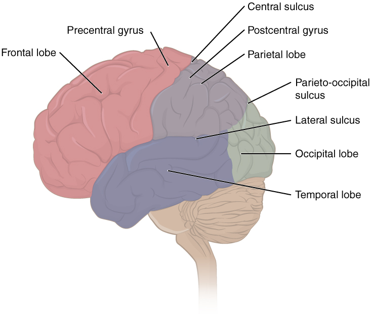
00:01 Good day. We´re gonna now discuss development of the cerebral cortex and the brain. 00:06 You may recall when we discussed the neural tube and the development of the brain stem, we had various internal portions of the neural tube that would expand to become the ventricles. 00:17 In the cerebral cortex, we have two extensions from the telencephalon that point out to the left and the right, and expand just tremendously. 00:26 This massive growth is going to be mirrored by a growth of the space inside to create the left and right lateral ventricles. 00:34 As the brain expands, the space inside them expands as well but remains connected to the third ventricle through the interventricular foramen. 00:43 So the growth of the brain is mirrored by the growth of its ventricles. 00:46 And we can see that as the brain´s cortexes take on their distinctive shape becoming the various lobes of the brain, it´s mirrored by the growth and extension of the ventricles within. 00:57 Now, growth of the brain is incredibly complex and the main points to take are that it develops in a similar way to the spinal cord and brain stem. 01:07 We have the ventricular zone close to the central canal which is going to become the ventricles. 01:12 Neurons and neuroepithelial cells in that area divide and start migrating more laterally and they´re gonna pass through a variety of spaces in the developing brain. 01:22 The subventricular zone, the intermediate zone, the cortical plane, and finally, the edge of the brain, the marginal zone. 01:30 But for the neurons to find their place in this process, they actually follow glial cells. 01:36 The supporting cells of the brain which were also derived from neuroepithelium have long extended processes that reach all the way from the medial to lateral aspects of the brain and these radial glial processes are actually there as a road for these neurons to take and they literally climb their way up these glial processes to find their appropriate place in the brain. 01:59 Now, the neurons in the brain develop a variety of layers and that´s what gives the distinctiveness to each region and as you may recall from neuroscience, the different Brodmann's areas are present because neurons migrate slightly differently in one region of the brain compared to the other. 02:14 Now, overall, the brain starts out growing and the cortexes expand but that only picks up more speed as time goes by. 02:24 From three months, to four months, to six months, the brain just tremendously expands and the various lobes start to become a little more evident and by the time we reach six months, the brain more or less looks like we´d expect it to with distinctive lobes starting to appear at that time. 02:42 The connection between the lobes called the corpus callosum becomes more and more stout as time goes by because neurons from one cortex are migrating to the other and vice versa. 02:55 So the corpus callosum is the nervous structure that allows one side of the brain to talk to the other. 03:00 Now, by six months, we have fairly distinct lobes of the brain and those lobes are going to be the frontal, parietal, occipital, temporal, and insular. 03:11 But early on, we don´t see a real clear distinction between those. 03:15 It´s not until near birth that the lobes and in particular, this gyri and sulci, the little tiny divisions of the brain are very, very evident. 03:24 So as we move from four months, to six months, to seven months, the brain is still relatively smooth and only the largest sulci and fissures are present. 03:34 Now, the brain needs to have space. 03:36 We have some real estate available for these neurons to inhabit. 03:40 So once the lobes have formed, the next thing it does are create those little ridges, the gyri is for the neurons to take up residence in. 03:48 Now, it´s not until about nine months that the brain looks pretty much like a brain we´d expect to see in an adult except in a smaller version. 03:58 The brain does continue to grow throughout childhood, adolescence, and into adulthood reaching its final stage at about the same time that the bones of the skull stop growing. 04:08 One thing that´s a little bit interesting is that we have a lobe of the brain that most people have not heard about. 04:13 It´s called the insular lobe and it´s located between the frontal, the parietal, and the temporal lobes. 04:18 It´s still present in adults but it´s gotten overgrown by the other lobes and to see it, we actually have to open up the lateral cerebral fissure and push the temporal lobe down, push the frontal and parietal lobes up to see that overgrown insular lobe. 04:34 Now, at the same time, the cerebellum and brain stem are developing and extending inferiorly from the cortex and at nine months, we more or less have a brain that looks typical. 04:44 Now, in addition to the brain itself, one cranial nerve extends off the cortex. 04:50 The others are coming from further down in the brain stem and the one that comes from the cortex is the olfactory nerve and its olfactory bulb. 04:57 Now, these develop as an extension of the telencephalon and we have a neural placode that hallows out to become a neural pit and create the nasal cavity. 05:08 As the nasal cavity is forming, these sensory nerve cells that are within it are going to innervate the mucosa and the upper third of the nasal cavity and connect to an extension of the telencephalon called the olfactory bulb. 05:23 Now, if you wanna be very technical, the olfactory nerves created in nerve one are just the nerves running from the nasal cavity to the olfactory bulb but most anatomists including myself refer to the entire assembly, olfactory bulb, tracts, and the nerves in the nasal cavity as cranial nerve one. 05:41 As the nose and mouth develop, these nerves get closer and closer into the mucosa, extend into the bulb and they´re present before bone is present in that area. 05:52 Therefore, we have bones developing in the nasal cavity and in the skull. 05:57 And the cribriform plate of the ethmoid bone has many, many tiny holes in it because it has to develop around the nerves that are already present. 06:07 And if you have any trauma to the head that sheers those nerves, you can wind up with loss of the sensation of smell. 06:13 So the olfactory bulb connects from the telencephalon and synapses with olfactory nerves coming from primary neurosensory cells in the nasal cavity. 06:25 Now, what things can go wrong in this process? If you recall, we discussed how the cranial and caudal neuropores close during early development neurulation and this allows the central nervous system to migrate completely into the mesoderm and take up residence inside the body. 06:44 If there´s a delay or outright failure of the cranial neuropore to close, it can create a variety of problems and their severity is matched by the impressive nomenclature that´s associated with them. 06:57 First, we can have a defect in the posterior aspect of the skull that allows the meninges and cerebrospinal fluid to balloon out. 07:05 This is gonna be called a meningocele because it has meninges, the dermodrin arachnoid mater pushing out from this bubble within the skull. 07:14 If we have a portion of the brain or cerebellum, so cortex or cerebellum pushing through a defect as well as the meninges and cerebral spinal fluid, this is called ameningoencephalocele. 07:28 And last but not least by a long shot is what happens when we have the meninges, cerebrospinal fluid, brain matter, and a portion of the internal ventricles herniating through a defect. 07:43 This is called a meningohydroencephalocele. 07:47 Now, the nomenclature sounds daunting but it really does describe what´s involved in the bubble. 07:52 If it´s meninges and CSF, it´s a meningocele. 07:55 If it´s meninges and part of the brain, it´s a meningoencephalocele and if it´s meninges, brain, and the internal ventricular system, then we have meningohydroencephalocele. 08:11 Very severe failure of the cranial neuropore to close results in a failure of the brain itself to form and this is going to be called anencephaly, failure of the head to develop. 08:21 This is absolutely incompatible with life outside the womb and because the neural tube didn´t form, the brain itself cannot form with this severity of a neuropore defect. 08:33 Now, other things that can go wrong in this process is failure of the right and left cortexes to fully separate from each other and this creates a spectrum of disease called holoprosencephaly. 08:45 So it´s located on the prosencephalon but there´s a fusion of it, hence, holoprosencephaly. 08:51 The least severe forms of this have slight narrowing of the midline structures of both the central nervous system and the face, and they can be relatively innocuous. 09:00 However, if the brain has not fully separated, we wind up with what´s called semilobar holopresencephaly where the right and left lobes of the brain are incompletely divided and it´s connected to a far greater degree than it should be. 09:16 In this image, you can see that the right and left cortex have not separated and the lateral ventricles are very, very open to one another in this brain. 09:25 The most severe form of holoprosencephaly is known as alobar holoprosencephaly where there´s simply no division of the right and left cortex from one another. 09:33 These brains appear somewhat mushroom shaped because they´re so close together on the midline, the eyes do not fully separate and we can have varying degrees of cyclopia or single eyed appearance. 09:45 So remember, that holoprosencephaly does not just affect the central nervous system but also, the structures of the face which we´ll discuss in the lecture on facial development. 09:55 Other problems that can happen in this process is that we can have failure of the brain to enlarge to the degree that we´d expect. 10:02 This is gonna case microcephaly, small headedness. 10:06 This is typically diagnosed when someone has a head that is two to three standard deviations below the norm. 10:14 It can happen alongside other conditions or sometimes, can happen just in isolation. 10:19 It is tied to a variety of problems including chromosomal abnormalities, different mutations, viral infections such as Zika, maternal malnutrition and other causes that go further on down the line of obscurity. 10:34 Microcephaly does result in developmental delays because there´s simply not as much neural tissue present and seizures are a common sequelae of it as well. 10:44 Thank you very much and I´ll see you on our next talk.
About the Lecture
The lecture Development of Cerebral Cortex by Peter Ward, PhD is from the course Development of the Nervous System, Head, and Neck. It contains the following chapters:
- Development of the Cerebral Cortex
- Neural Tube Defects in the Skull
Included Quiz Questions
Supporting glial cells are derived from which embryonic layer?
- Neuroepithelium
- Surface ectoderm
- Neural crest
- Intermediate mesoderm
- Lateral plate mesoderm
What brain structure connects the left and right cortex?
- Corpus callosum
- Central sulcus
- Calcarine sulcus
- Lateral sulcus
- Cuneus
The olfactory bulb is the extension of what embryonic brain vesicle?
- Telencephalon
- Mesencephalon
- Metencephalon
- Myelencephalon
- Diencephalon
What condition arises when there is a defect in the skull containing the meninges, CSF, ventricular system, and a portion of the cerebellum?
- Meningohydroencephalocele
- Meningocele
- Meningoencephalocele
- Tonsillar herniation
- Dandy-Walker syndrome
Failure of cerebral hemispheres and lateral ventricles to separate into left and right parts leads to what condition?
- Holoprosencephaly
- Anencephaly
- Microcephaly
- Meningoencephalocoele
- Hydrocephalus
Microcephaly (a head circumference between 2–3 standard deviations below the norm) is associated with all of the following causes EXCEPT?
- Complete failure of the cranial neuropore to close
- Chromosomal abnormalities
- Gene mutations
- Viral infections
- Maternal malnutrition
Customer reviews
5,0 of 5 stars
| 5 Stars |
|
5 |
| 4 Stars |
|
0 |
| 3 Stars |
|
0 |
| 2 Stars |
|
0 |
| 1 Star |
|
0 |


