Playlist
Show Playlist
Hide Playlist
Dermatomyositis

-
Slides Inflammatory Myopathies.pdf
-
Download Lecture Overview
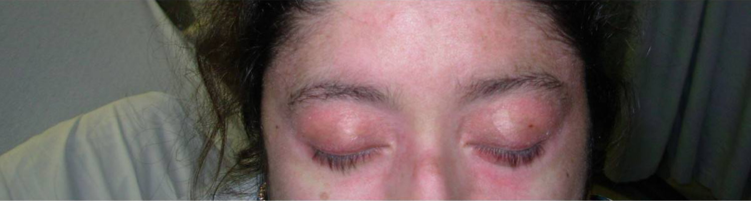
00:00 So let's now move to dermatomyositis, that second major inflammatory myopathy and let's think about some of the similarities to polymyositis and some of the differences. 00:12 Dermatomyositis also has a subacute onset. Patients present over weeks, and sometimes days, with weakness. The distribution is similar to polymyositis. Patients present with proximal weakness, difficulty getting upstairs and out of chairs, reaching up to the top cabinets. It's often symmetric. It should also be painless. And importantly, the distribution is proximal more than distal. We don't see prominent sensory findings. The sensory exam should be normal. 00:42 And reflexes should be normal to decreased. Importantly, with dermatomyositis it looks the same as polymyositis, except we see a rash. That rash may predate the weakness. In rare cases, the rash may show up after the weakness. But that rash is critical in differentiating between polymyositis and dermatomyositis. So, there's 3 types of skin findings that I would like for you to understand and know about when we're evaluating patients for a diagnosis of dermatomyositis. The first are Gottron’s papules and we saw these also in the last slide. This is a violaceous, a violet or purple hued papular rash. It's a raised rash that you can feel that typically forms over the knuckles as you can see here. We can see the heliotrope rash, which again is a violaceous, violet colored rash on the malar aspect of the face. And the third is the shawl rash, which you see on the chest area here. And the presence of that rash should really guide us and point us in the direction of a diagnosis of dermatomyositis over polymyositis. What's the work up? Well, once we have localized to the muscle and we're concerned about the rash, we need to prove this is inflammatory and that's an inflammatory myopathy. So again, we check the CK, and a CK in the thousands is consistent with the diagnosis of dermatomyositis. We can check more nonspecific muscle enzymes but that's not required particularly when we see elevated CK. The EMG can also be helpful and again we see myopathic units. There are small units, there are short duration, there's less muscle that's being activated and we can commonly see spontaneous activity. Those muscles are firing on their own because of inflammation around the muscle that's impairing signaling across the neuromuscular junction. And again, muscle biopsy can be helpful in cases where the diagnosis is unclear but it's not required to make a diagnosis of dermatomyositis. The treatment of choice is again corticosteroids similar to polymyositis and prednisone is typically the corticosteroid that would be selected. And this is another example of that shawl rash that we talked about on the last slide. What about the pathophysiology of dermatomyositis? What's going on at the level of the muscle to cause this condition? Well, again, it's inflammatory. So we're going to talk about the immune system, but the type of inflammation, the location of the inflammation is different and is important in a muscle biopsy in differentiating between the 2 conditions, poly- and dermatomyositis. Here we see that one of the drivers of dermatomyositis is complement activation. And we see in the systemic circulation, complement is activated and there's cytokine release and this brings a host of immune cells into and around the blood vessels of the muscle. We see activation of macrophages which travel into the interstitium around the perivascular area. We see activation of T-cells, which can communicate to B-cells, and bring B-cells and T-cells into that perivascular niche of the muscle. 03:35 And then there are other types of T-cells that traffic to and move into the perivascular area around the muscle. Ultimately, all of this inflammation within the interstitium drives cytokine release and we see that that normal muscle fiber, that normal fascicle where we have myocytes and capillaries in between the individual muscle fibers becomes damaged. We see capillary destruction. This is a perivascular inflammation and we see perifascicular atrophy. 04:04 And that perivascular and perifascicular atrophy are critical differentiators between dermatomyositis and polymyositis as you see here. Ultimately, we see lumen enlargement of the blood vessels, degeneration necrosis and atrophic myocytes and this is what's causing the patients to be weak. So what do we see on the biopsy? Well, again, what we're looking at here is the muscle. The large pink areas are muscle fibers and in between that is the interstitium and perimysial and endomysial tissue around it. Here we see that in between each of the muscle fibers, we see a lot of inflammation, those purple lymphocytes infiltrating into each muscle fiber and resulting in degeneration. And there's 2 key findings that I would like for you to take away from the muscle biopsy of dermatomyositis. We see perimysial and perivascular inflammation. And that's a key differentiator between polymyositis.
About the Lecture
The lecture Dermatomyositis by Roy Strowd, MD is from the course Acquired Neuromuscular Diseases.
Included Quiz Questions
A patient has proximal muscle weakness, a heliotrope rash, and Gottron papules. What is the one of the underlying drivers in the pathogenesis of the condition?
- Complement activation
- Intrafascicular infiltration of CD8+ T cells
- Statin-induced damage
- Trinucleotide expansion
- Frameshift mutation
Which of the following tests may be helpful in confirming suspected dermatomyositis?
- Muscle biopsy
- Serum ANA titer
- Skin biopsy
- Serum anti-Jo-1 titer
- Nerve conduction studies
Dermatomyositis is associated with which of the following symptoms?
- Erythema of the upper back, posterior neck, and shoulders ("shawl rash")
- Discoloration of the digits due to vasospasm
- Fibrosis of the lung
- Permanent vision loss
- Point tenderness
What will a muscle biopsy show from a patient suffering from dermatomyositis?
- Perimysial and perivascular inflammation
- Endomysial inflammation
- Fatty replacement
- Intramuscular vacuoles
- Normal findings
Author of lecture Dermatomyositis

Roy Strowd, MD
Customer reviews
5,0 of 5 stars
| 5 Stars |
|
5 |
| 4 Stars |
|
0 |
| 3 Stars |
|
0 |
| 2 Stars |
|
0 |
| 1 Star |
|
0 |
