Playlist
Show Playlist
Hide Playlist
Cutaneous Tuberculosis in Darker Skin
-
Slides Cutaneous Tuberculosis in Darker Skin.pdf
-
Download Lecture Overview
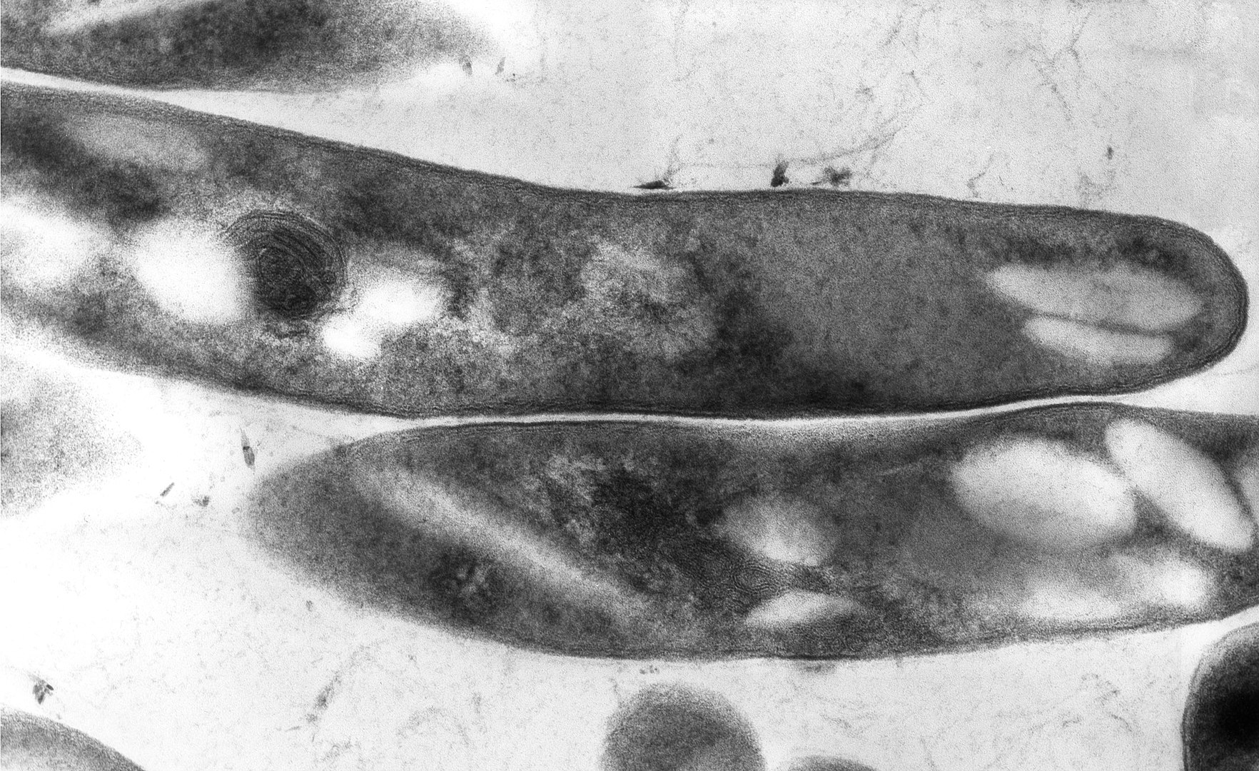
00:01 Welcome to our lecture on cutaneous tuberculosis or skin TB. 00:07 Cutaneous tuberculosis is invasion of the skin by Mycobacterium tuberculosis complex. It's relatively uncommon, but it does occur. Extrapulmonary TB constitutes 15% of all cases, and cutaneous lesions account for less than 2% of all extrapulmonary manifestations. So we know that the most common type of TB is pulmonary TB. So now let's look at the etiology of cutaneous TB. This is caused by gram positive rod shaped bacilli as listed. 00:52 So how do we classify cutaneous TB. 00:56 There's true cutaneous tuberculosis one. 00:59 Secondly, the tuberculids. 01:02 The tuberculids are a result of hypersensitivity reaction to mycobacteria that is not detectable, whereas in true cutaneous tuberculosis there's mycobacteria which can be identified in the areas of cutaneous TB. 01:18 So one gets contiguous spread or autoinoculation, and it can present as scrofulodema or TB cutis or orificialis. If there's hematogenous spread to the skin, you can get cutaneous TB presenting as lupus vulgaris, miliary TB, or metastatic tuberculosis abscess. Through inoculation from exogenous source, then you get primary inoculation which presents as TB verrucosa cutis or TB chancre. 01:57 Looking at now the tuberculids remember we mentioned that tuberculids is an ID reaction. It's a hypersensitivity reaction to the TB, but you cannot identify the TB in the lesion. 02:08 So what are the types of tuberculids? Erythema nodosum which is the most common presentation. 02:15 Erythema in duratom of basin or just erythema in duratom. 02:19 And what we call papular necrotic tuberculoid or PNT. 02:24 Lichen scrofulaceum, which is more common in children. 02:28 Now let's now focus on the individual types of cutaneous TB. 02:32 We want to start with scrofulo derma. 02:34 This results from direct extension of an underlying TB focus could be a regional lymph node or infected bone or joint. 02:42 And then you get spread to the surface. 02:46 And this presents with firm, painless lesions that eventually ulcerate with a granular base. It may heal even without treatment, but it takes years and leaves unsightly scars. 02:59 So if again focus on scrofuloderma and look at the differential of scrofuloderma. Remember we're talking about the different types of TB. 03:08 We're starting with scrofuloderma and its differentials. 03:11 So the differential of scrofuloderma is bacterial abscess a condition called hidradenitis suppurativa which has become quite common in the recent years and common in patients who are overweight. 03:26 And the other differential is gummatous syphilis. 03:30 Moving on now to the second type it's lupus vulgaris. 03:36 Lupus vulgaris, you get sharply defined grayish brown nodules with a gelatinous consistency. 03:42 You don't see the erythema. 03:44 Erythema that's classical in white patients. 03:47 It affects primarily the head and neck and persists for years, leading to disfigurement and skin cancer in some cases. 03:58 The differential of lupus vulgaris includes other forms of cutaneous TB, for example, tuberculosis verrucosa cutis. 04:08 The second differential is deep fungal infections, for example histoplasmosis and of course lepromatous leprosy can be also confused with lupus vulgaris. 04:25 Leishmaniasis is another condition which is seen commonly in South America, and it's a big differential in that area where it is endemic for lupus vulgaris. 04:38 Other differentials for lupus vulgaris include sarcoidosis and Wegener's granulomatosis. 04:48 So moving on to tuberculosis. 04:52 This is a hypersensitivity reaction due to circulating Mycobacterium tuberculosis or its antigens. 05:02 The first one that we're going to talk about is erythema nodosum. 05:06 It is the most common tuberculoid, and it presents with bilateral tender, erythematous subcutaneous nodules ranging from 3 to 20cm. 05:17 It tends to occur on the arms, knees, lower legs, sometimes on the face and neck, but this is very rare, if it when it affects the face and neck. 05:29 So what are the differentials of erythema nodosum erythema induratum of basin which is another type of Id reaction or hypersensitivity reaction. Cutaneous polyarteritis nodosa, which is a connective tissue disorder, is another differential for erythema nodosum, and of course superficial thrombophlebitis is a differential for erythema nodosum. The next type of id reaction is erythema induratum of bazin. 06:03 Here one gets recurrent nodules or lumps, and they tend to be bigger than those of erythema nodosum. 06:11 Same year it tends to occur in lower legs, particularly young women. 06:17 It may ulcerate and scar, and the differential involves erythema nodosum and cutaneous polyarteritis nodosa, and of course, cutaneous lymphoma. The next step of tuberculin is what we call P N T, papulo necrotic tuberculin. 06:36 In these patients, you see multiple recurrent hypopigmented crusted papules. 06:43 The papules can become pustular or necrotic, resulting in small ulcers and scars after healing. 06:53 Typically heals with varioli form, chickenpox like scarring after about six weeks. And it tends to occur on the upper limbs and on the lower limbs. The differential diagnosis includes leukocytoclastic vasculitis, papular eczema, and prurigo simplex. The last one, lichen scrofulosorum, is another id reaction. 07:19 It's a lichenoid eruption of minute papules, hence the name lichen and papules are usually asymptomatic. 07:27 They are closely grouped and their skin colored to brown color. 07:33 The differential of lichen scrofulosorum is lichen nitidus, which is usually asymptomatic, and another one is keratosis pilaris, which is quite common and sometimes associated with atopic eczema. So what are some of the complications of these conditions? Dissemination is very rare and one m ay get scarring. How do we diagnose the skin, cutaneous TB? The history and physical examination is important, and immunologic tests are done, for example, the mantoux or tuberculin skin test, which is a delayed hypersensitivity reaction, is positive in patients with skin TB. 08:17 We also offer the interferon gamma release assays to see whether the patient has been exposed to TB. 08:25 Mycobacterial culture remains the gold standard for non-tuberculous lesions, but in the id reaction that you're not going to find anything because it's a hypersensitivity reaction. 08:39 The skin biopsy is also an important investigation that we have to do where we suspect cutaneous TB. 08:48 And it's also important to trace the contacts of family members of patients with TB. 08:58 What about management? The treatment of TB is the standard anti-TB therapy which includes rifampicin, isoniazid, pyrazinamide and ethambutol . 09:09 And we use a nice mnemonic RIPE therapy so you don't forget. 09:14 So there's two phases. 09:16 The first phase is intensive phase where there's two months of all four anti-TB treatment. This is then followed by the maintenance phase which includes four months of only rifampicin and isoniazid. And of course it's important in black patients because of the scarring and post-inflammatory hyperpigmentation to ensure that you offer treatment for patients for a holistic approach to anti TB medication.
About the Lecture
The lecture Cutaneous Tuberculosis in Darker Skin by Ncoza Dlova is from the course Bacterial Skin Infections in Patients with Darker Skin.
Included Quiz Questions
Which of the following is the key difference between true cutaneous tuberculosis and tuberculids?
- Mycobacteria can be identified in lesions of true cutaneous TB but not in tuberculids
- Tuberculids affect only the dermis while true cutaneous TB affects subcutaneous tissue
- True cutaneous TB is always associated with pulmonary TB while tuberculids are not
- Tuberculids respond to standard anti-TB therapy while true cutaneous TB does not
- True cutaneous TB is painless while tuberculids are typically painful
Which form of cutaneous tuberculosis results from hematogenous spread to the skin?
- Lupus vulgaris
- Tuberculosis verrucosa cutis
- Tuberculosis chancre
- Scrofuloderma
- Primary inoculation tuberculosis
A patient presents with recurrent multiple lower extremity dark papular lesions, some of which have become pustular. Pathology showed tuberculoid granulomatous inflammation but smears and culture were negative. What is the diagnosis?
- Papular necrotic tuberculid
- Lichen scrofuloderma
- Erythema induratum of Bazin
- Lupus vulgaris
- Tuberculosis verrucosa cutis
Which test remains the gold standard for diagnosing non-tuberculid cutaneous lesions?
- Mycobacterial culture
- Mantoux test
- Interferon gamma release assay
- Skin biopsy histopathology
- Polymerase chain reaction
Which characteristic feature is associated with lupus vulgaris?
- Sharply defined grayish-brown nodules with gelatinous consistency
- Firm painless lesions that eventually ulcerate with granular base
- Multiple recurrent hypopigmented crusted papules
- Bilateral tender erythematous subcutaneous nodules
- Lichenoid eruption of minute asymptomatic papules
Customer reviews
5,0 of 5 stars
| 5 Stars |
|
5 |
| 4 Stars |
|
0 |
| 3 Stars |
|
0 |
| 2 Stars |
|
0 |
| 1 Star |
|
0 |


