Playlist
Show Playlist
Hide Playlist
Cranial Nerve VII: Facial Nerve

-
Slides 8 CranialNerves 2 BrainAndNervousSystem.pdf
-
Reference List Anatomy.pdf
-
Download Lecture Overview
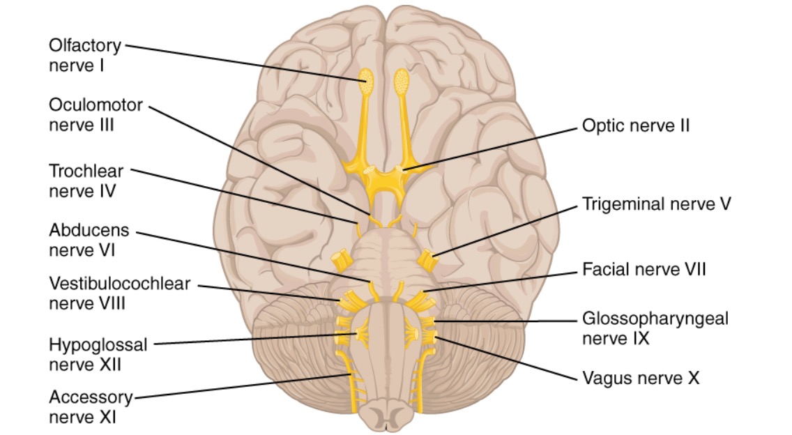
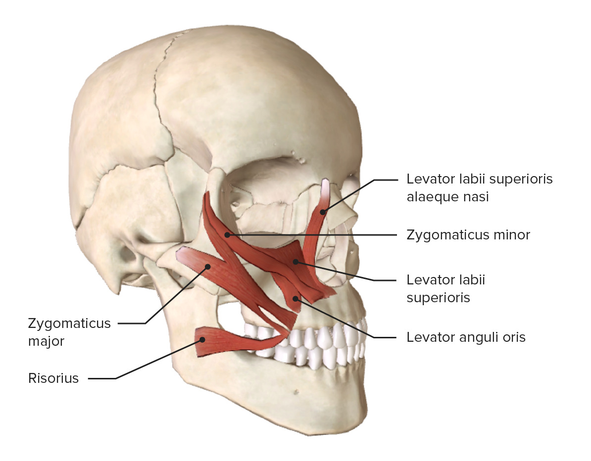
00:00 We’ll take a look at each one of these cranial nerves starting with the facial nerve. First thing for you to understand is what are the functional components that are conveyed through the facial nerve fibers. 00:17 I want you to understand the functional components that are associated with this nerve. This nerve will convey general somatic afferent nerve fibers. It also carries special afferent nerve fibers. Thirdly, it carries general visceral efferent nerve fibers. Then lastly, it has branchial efferent nerve fibers because the facial nerve during embryology or development supply the second pharyngeal arch. The facial nerve enters through the internal acoustic meatus and exits through stylomastoid mastoid foramen. 01:03 Next, we’ll take a look at the distribution of each one of these functional components. 01:10 I’ll have you begin with an understanding of the general somatic afferents. 01:16 These are going to innervate the external acoustic meatus and the deeper parts of the auricle or the outer part of the ear. The facial nerve also conveys special afferent nerve fibers. 01:39 In this case, these nerve fibers are conveying taste from the anterior two-thirds of the tongue and you can see the anterior aspect of the tongue here shaded in red. With respect to general visceral efferents, the facial nerve is going to innervate the lacrimal gland through parasympathetics. It also innervates the submandibular and sublingual salivary glands. It’s also responsible for innervating the mucous membranes of the nasal cavity and the area of the hard and soft palates. The branchial efferent component, and this is the last functional component, is going to innervate muscles that were derived from the second pharyngeal arch. This would include muscles of facial expression that have now been shaded in red. It’s also responsible for innervating the stapedius, a small muscle that’s found in the middle ear that attaches to the stapes. The digastric muscle, specifically the posterior belly is innervated by the facial nerve. The last muscle derived from the second pharyngeal arch to be innervated by the facial nerve is your stylohyoid muscle shown in through here. A clinical consideration with the facial nerve is that of Bell’s palsy. This is a palsy of the seventh cranial nerve. Individuals that have Bell’s palsy have some symptoms that are associated with the functional components of the facial nerve. 03:56 One symptom would be weakness to paralysis of the facial muscles. Drooling is a symptom associated with Bell’s palsy in that the sphincter around the mouth, the orbicularis oris is partially paralyzed or weak. 04:19 Drooping of the lower eyelid is another symptom. Individuals with Bell’s palsy may complain of sensitivity to sound, hyperacusis. This would occur if there’s paralysis to the stapes muscle. Because the facial nerve also conveys gustation or taste, individuals may complain of impairment or even loss of taste. 04:57 The cause of Bell’s palsy is a viral infection. It can be treated with antiviral agents. To reduce the inflammation associated with the infection, steroids can also be administered. They’re usually effective as well.
About the Lecture
The lecture Cranial Nerve VII: Facial Nerve by Craig Canby, PhD is from the course 12 Cranial Nerves and Their Functions.
Included Quiz Questions
Which statement about cranial nerve VII is INCORRECT?
- It innervates the parotid gland.
- It is responsible for the secretions of the submandibular salivary glands.
- It innervates the muscles of facial expression.
- It carries general somatic afferents from the external acoustic meatus.
- It innervates the sublingual salivary glands.
Which condition would result if loss of innervation to the stapedius muscle occurred?
- Hyperacusis
- Drooling of saliva
- Loss of the corneal reflex
- Difficulty swallowing
- Drooping of the lower eyelid
Which condition could result if Bell’s palsy occurred?
- Sensitivity to sound
- Hearing loss
- Fixed and dilated pupil
- Loss of pain sensation from the anterior two-thirds of the tongue
- Spastic paralysis of muscles of mastication
Author of lecture Cranial Nerve VII: Facial Nerve

Craig Canby, PhD
Customer reviews
2,6 of 5 stars
| 5 Stars |
|
2 |
| 4 Stars |
|
0 |
| 3 Stars |
|
1 |
| 2 Stars |
|
1 |
| 1 Star |
|
3 |
Se requiere mas infromación, incluyendo aquellas paralisis qu ese pueden dar por un tumor.
covered very well contains the information needed for step in med school
good explanation of the components in the facial nerve. Thanks
It`s almost nothing in this lecture. We need some raports and traiects of the nerves.
