Playlist
Show Playlist
Hide Playlist
Colonic Polyps with Case
-
Slides Large Intestine.pdf
-
Reference List Gastroenterology.pdf
-
Download Lecture Overview
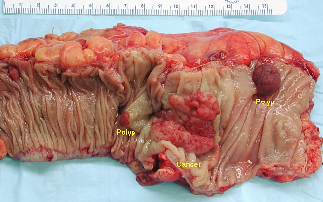
00:01 We'll move on to our next case. 00:03 We have a 59-year-old woman who undergoes a routine screening called colonoscopy. 00:08 On colonoscopy, she is found to have a 15 mm polyp in the descending colon and no other lesions. 00:16 The polyp is removed. 00:18 Pathology shows a villous adenoatous polyp. 00:21 She asks how these findings relate to her risk of developing colorectal cancer. 00:27 So how should she be counseled and is this a lesion with malignancy potential? Let's go to some key features here. 00:35 So, she has a large polyp which is probably a high risk. 00:40 And she has a villous adenoma on her pathology. 00:43 We'll talk a bit more about what that means. 00:48 So we categorize colonic polyps in 2 different ways. 00:52 First, their gross appearance on endoscopy. 00:55 And second, by histology. 00:57 So first let's focus on their appearance You may have a sessile polyp, which is when you have a polyp and majority of its base is attached to the colonic wall as shown here. 01:09 You may also have a pedunculated polyp. 01:12 This is when the polyp is attached by a short stalk to the colonic wall. 01:19 And lastly, you may have a flat polyp. 01:21 These are as you can imagine, some of the most difficult polyps you detect. 01:25 as there is not much to distinguish it from the rest of the colonic wall. 01:31 Next, we also categorize polyps based on their appearance on histology. 01:36 So you may have an adenomatous polyp which is usually just a benign growth of tubular glands that grow from the colonic wall. 01:45 You may have a serrated polyp. 01:47 which is a typical sawtooth like appearance of the glands on histology. 01:52 And you may have other types of histologic polyps including hamartomas and other things that we will not cover here. 02:01 So some higher risk features of polyps include larger size, so specifically anything larger than or equal to 10 millimeters. 02:10 The histologic type of anomalous polyp tends to be the highest risk. 02:15 Those that have high degrees of dysplasia as seen on histology are also at elevated risk. 02:23 And lastly, a sessile shape can also be a high risk feature. 02:28 So if you have any pf these high risk features, these polyps should be removed. 02:33 And patients who have those should be surveilled with colonoscopy at more frequent intervals than the normal. 02:41 So now let's talk a bit more in detail about adenomatous polyps. 02:46 Within that broad category of adenomatous polyps, there are different types of histology that are more worrisome than others. 02:53 So first is a tubular adenoma. 02:56 This is when the majority of the polyp is made up of branching tubules as shown here. 03:01 It is the most common, about 80% of all adenomas are this type. 03:07 You may also have a tubulovillous adenomatoma which is a mixed picture between tubular and villous. 03:15 This makes up about 15% of all adenomas. 03:19 The last category is a villous adenoma. 03:23 In this, you have the majority of long glands that then extend to the center. 03:28 And this again, is about 15% of adenomas. 03:31 This is important because as you go from tubular, to tubulovillous and lastly to villous, you have increasing risk of malignancy. 03:39 So villous adenoma is the highest risk of progressing to malignancy. 03:46 So treatment of colonic polyps typically consist of complete removal of the polyp and then considering surveillance colonoscopy on follow up. 03:55 How often you do surveillance colonoscopy depends on the number of factors including: size, the number of polyps that were found, and the histology of the polyps. 04:07 So now we can return to our case. 04:09 We had a 59-year-old woman undergoing routine screening colonoscopy. 04:14 She has a large polyp that's greater than 10 mm in size - so that is a high risk feature. 04:19 In addition, she has villous adenoma histology which is as we know now, also at high risk for dysplasia. 04:28 So how should she be counseled and is this a lesion with malignancy potential? We now know that based on the size and the histology of her polyp, this is now a high-risk polyp that was removed so she now needs more frequent surveillance colonoscopy, you might consider repeating one in 3 years.
About the Lecture
The lecture Colonic Polyps with Case by Kelley Chuang, MD is from the course Disorders of the Small and Large Intestines.
Included Quiz Questions
Which of the following adenomatous polyps has the highest risk for malignancy?
- Villous adenoma
- Tubular adenoma
- Tubulovillous adenoma
- Hamartoma
- Serrated polyp
Which of the following is the best next step after the removal of a high-risk colon polyp?
- Colonoscopy surveillance
- Abdominal CT surveillance
- Abdominal X-ray surveillance
- Sigmoidoscopy surveillance
- Abdominal ultrasonography surveillance
Customer reviews
5,0 of 5 stars
| 5 Stars |
|
1 |
| 4 Stars |
|
0 |
| 3 Stars |
|
0 |
| 2 Stars |
|
0 |
| 1 Star |
|
0 |
All of her lectures are awesome , she should make more lectures , wonderful teacher


