Playlist
Show Playlist
Hide Playlist
Cellulitis in Darker Skin

-
Slides Cellulitis in Darker Skin.pdf
-
Download Lecture Overview
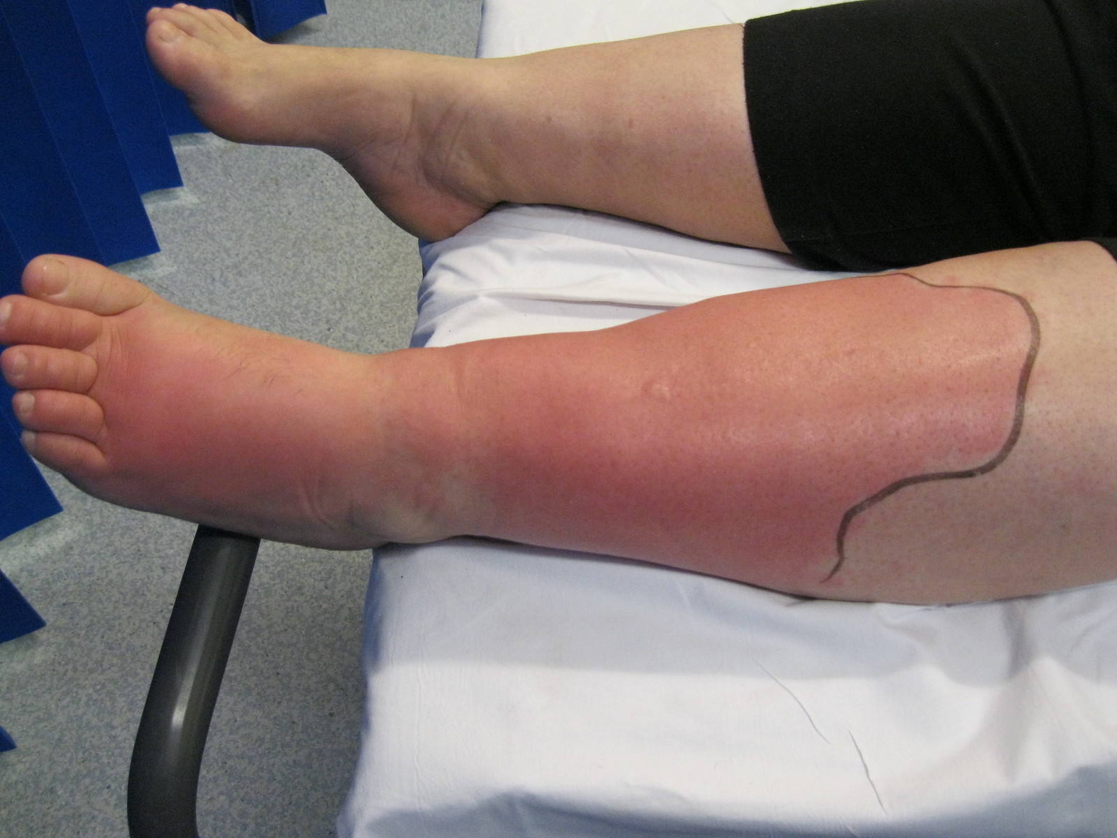
00:01 Welcome to a lecture on cellulitis. 00:05 How do we define cellulitis? It's an acute infective inflammatory condition of the dermis and subcutaneous tissue. It's often a complication of wounds, ulcers or other skin conditions. 00:21 It's most common in middle aged and older adults. 00:27 And some of the risk factors to cellulitis include skin barrier disruption due to trauma, skin inflammation, edema, obesity, and immunosuppression due to HIV or chemotherapy or cancer. Sometimes, preexisting skin infection can also predispose to cellulitis, as cellulitis is most commonly caused by group A beta hemolytic streptococcus, but it can also be caused by staph aureus and sometimes gram negative aerobic bacilli. 01:07 Now let's quickly go over pathophysiology of cellulitis. 01:11 In order for cellulitis to develop, there needs to be a breach in the skin. 01:16 This could be in the form of a fissure, a cut, laceration, insect bite, or any other mechanism that breaks the barrier of the skin. This is how strep pyogenes, or any other bacterial infection invades deeper layers of the skin. 01:32 Once in the epidermis and dermis, it starts proliferating and producing different exotoxins that induce inflammation and tissue destruction, thus eventually leading to cellulitis. 01:45 So that's the pathophysiology of cellulitis. 01:51 The most common site of involvement are the lower extremities, and it's almost always nearly unilateral. 01:59 If a patient presents with bilateral involvement you have to consider alternative causes. Clinically it presents with erythema or a grayish dark hue in skin of color with edema and a warm feeling to touch. 02:18 It has a poorly defined border, and in severe cases blisters may also be seen, and this is called cellulitis bullosa. 02:29 Fever and other systemic manifestations. 02:32 Manifestations may also be present. 02:34 The complications of cellulitis involve lymphangitis, gangrene and septicemia. 02:43 The diagnosis is based on clinical history and physical examination. 02:47 A swab needs to be done when necessary, and serologic testing for beta hemolytic strep in cases of recurrent cellulitis is advisable. 03:01 What are some of the differential diagnosis of cellulitis? Firstly, necrotizing fasciitis. 03:07 In this condition we get involvement of deeper tissue, for example the muscle fascia and overlying subcutaneous fat. 03:16 Erysipelas is another differential for cellulitis. 03:19 Here we have a clear demarcation between involved and an involved tissue, which is totally different from cellulitis, where there is no clear demarcation. Contact dermatitis is another differential, but this is usually associated with pruritis and generally limited to the site of the contact with the allergen. 03:42 The management of cellulitis if it's mild, oral antibiotics are given, particularly the penicillins. 03:51 Tetracyclines would be recommended if there's MRSA is cultured or suspected. 03:58 In severe cases of cellulitis high doses of intravenous antibiotics, for example vancomycin, vancomycin, and cefepime, may be recommended. The duration of treatment depends on the severity and clinical response, but usually takes about 5 to 6 days. 04:18 Generally, we also advise patients to encourage lymph movement and gentle exercise, which may reduce lymphedema if present.
About the Lecture
The lecture Cellulitis in Darker Skin by Ncoza Dlova is from the course Bacterial Skin Infections in Patients with Darker Skin.
Included Quiz Questions
What is a characteristic feature of the borders in cellulitis compared to erysipelas?
- Poorly defined borders
- Clearly demarcated borders
- Vesicular borders
- Hyperpigmented borders
- Satellite lesions at the borders
Which clinical feature should prompt consideration of an alternative diagnosis to cellulitis?
- Bilateral lower extremity involvement
- Unilateral lower extremity involvement
- Presence of local warmth and edema
- Poor response to first-generation cephalosporins
- Development of fever within 48 hours
Author of lecture Cellulitis in Darker Skin

Ncoza Dlova
Customer reviews
5,0 of 5 stars
| 5 Stars |
|
5 |
| 4 Stars |
|
0 |
| 3 Stars |
|
0 |
| 2 Stars |
|
0 |
| 1 Star |
|
0 |
