Playlist
Show Playlist
Hide Playlist
Cells of the Lymphatic System

-
Slides 11 Human Organ Systems Meyer.pdf
-
Reference List Histology.pdf
-
Download Lecture Overview
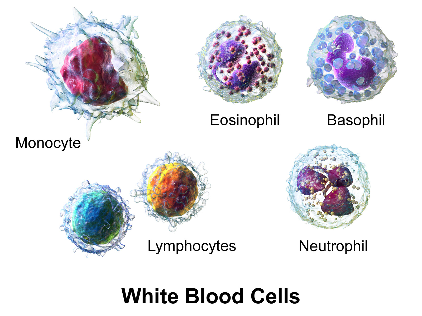
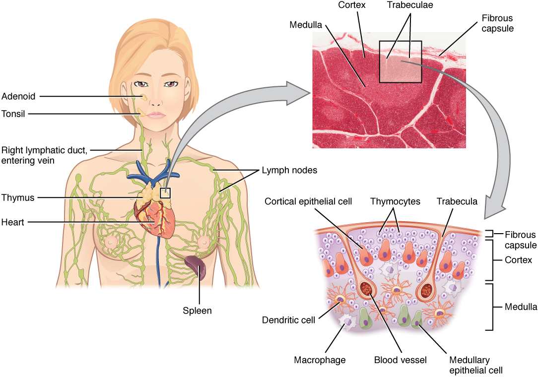
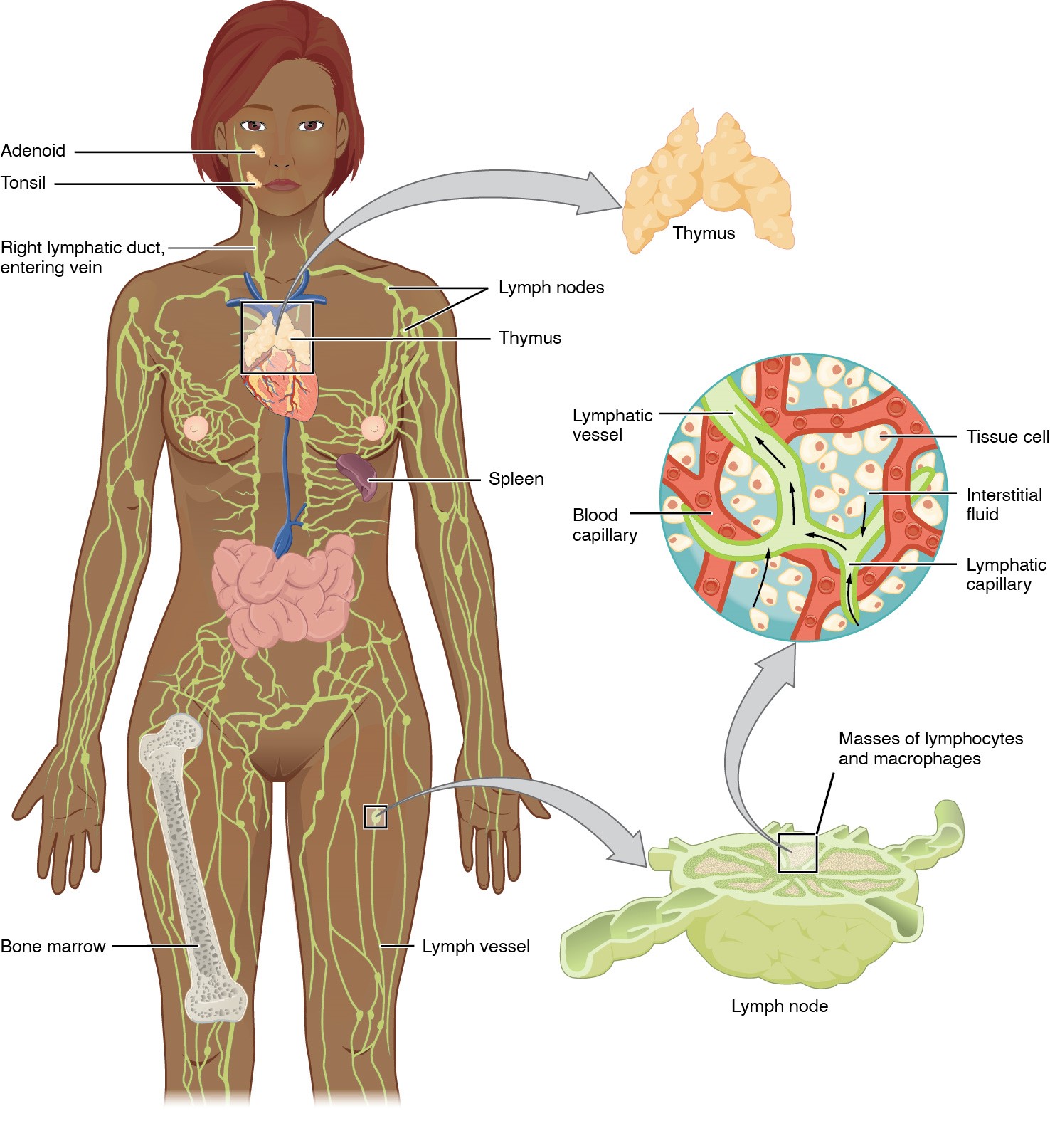
00:00 When we look at the cells that are involved in immune reactions, we can classify them into two different classes. 00:09 First of all, there are the lymphocytes, and then there are the accessory cells or helper cells, antigen presenting cells they’re often referred to. The lymphocytes circulate around the body in the blood, and they leave the blood in various locations by means I’ll describe throughout this lecture. And then they can go through the tissues undergoing surveillance on sensory duty to detect an antigen. And these lymphocytes fall into two different classes. B cells which differentiate in bone-marrow and a bone-marrow derived, and the T cells which although they derive from bone-marrow, they need to go to the thymus gland where they’re educated to detect and respond to their antigens that they are trying to detect. 01:03 If you remove the thymus from an experimental animal, they don’t develop T cells. 01:11 And for that reason, the T cells are thymus-dependent on their education, T for thymus, T for T cells. The B cells were allegedly described in the bursa of Fabricius, which is a little gland they discovered in chicken embryos in the cloacal region. And they are found to contain lymphocytes and removal of this bursa of Fabricius experimentally result in no antibodies being produced in the animal. So they are called B for B cells, B for the bursa of Fabricius. So that’s where the terms B and T come from. And within the T cells, there are two categories, helper T cells, cytolytic T cells, and also other sorts of T cells. 02:02 The accessory cells are macrophages, dendritic cells, and follicular dendritic cells, which I’ll refer to throughout this lecture. When we look at the B cells, the T cells, natural killer cells, they are the key players. But as I alerted to you in the previous slide, these other four groups of cells play a very important role. Usually, lymphoid tissues, organs such as the lymph node and spleen, they are dominated by a very, very elaborate spongy network that consist of reticular cells. These reticular cells secrete reticular fibres or collagen type III, and they wrap around those fibres and form this cobwebby spongy network right throughout the organ. And they express little flags on their surface to attract in all the four cell types that you see listed in this slide. The monocytes, macrophages, some of the blood cells, some of the white blood cells moving to these spaces, attracted by the reticular cells. Reticular cells also hone in dendritic cells. It’s important to understand that Langerhans cells are dendritic cells, but follicular dendritic cells are not. 03:31 They’re not derived from the stem cell. They live in lymph nodes which we’ll see in a moment. 03:38 And the epithelioreticular cell is the word I’ll describe later on that needs a little bit of elaboration because again, epithelioreticular cells are confined to the thymus. They are the supporting cell in the thymus. They’re not reticular cells. They don’t secrete reticular fibres. They’re epithelial cells. So perhaps, the name needs correction. 04:05 One way in which we find the histology of lymphoids difficult in some respect is that, there are many many different sorts of lymphocytes, and they can be identified using certain markers by immunohistochemical staining using monoclonal antibodies. But if you don’t use these stains, it’s very hard to differentiate different sorts of cells. They express these CD molecules called cluster of differentiation markers. And various lymphocytes express certain sorts of these CD molecules. Some cells express particular CD molecules for their entire lifespan. 04:54 Others only express these CD molecules at certain stage of their differentiation, or when they’re activated. And based on those three different criteria, immunologists and medical laboratory scientists can use histochemical staining, immunohistochemical staining using monoclonal antibodies that I mentioned a moment ago to actually identify and differentiate these different sorts of lymphocytes.
About the Lecture
The lecture Cells of the Lymphatic System by Geoffrey Meyer, PhD is from the course Lymphoid Histology.
Included Quiz Questions
Which of the following is INCORRECT?
- T-cell differentiation takes place mainly in the lymph nodes.
- Reticular cells produce and maintain the thin networks of fibers that are the framework for most lymphoid organs.
- In the thymus, epithelioreticular cells form the structural framework.
- Cluster of differentiation molecules are unique cell surface molecules and can be visualized by immunohistochemical staining methods using monoclonal antibodies.
- Some B cells differentiate and give rise to cells that secrete antibodies.
Where do T cells generally mature?
- Thymus
- Thyroid
- Spleen
- Bone marrow
- Lymph nodes
Which of the following is NOT a lymphocyte?
- Follicular dendritic cell
- B cell
- Natural killer cell
- Helper T cell
- Cytotoxic T cell
Reticular fibers, secreted by reticular cells, are composed of which of the following types of collagen?
- Type 3
- Type 2
- Type 1
- Type 4
- Type 5
Which cluster of differentiation (CD) is most characteristically associated with helper T cells?
- CD4
- CD3
- CD2
- CD20
- CD8
Author of lecture Cells of the Lymphatic System

Geoffrey Meyer, PhD
Customer reviews
5,0 of 5 stars
| 5 Stars |
|
1 |
| 4 Stars |
|
0 |
| 3 Stars |
|
0 |
| 2 Stars |
|
0 |
| 1 Star |
|
0 |
Excellent knowledge Amazing lecturer, understands how to contextualize different concepts and aspects of the knowledge. Understand how to BUILD the knowledge on itself.
