Playlist
Show Playlist
Hide Playlist
Case: 51-year-old with Left Hand Numbness

-
Slides Multiple Sclerosis Inflammatory Disorders of the CNS .pdf
-
Download Lecture Overview
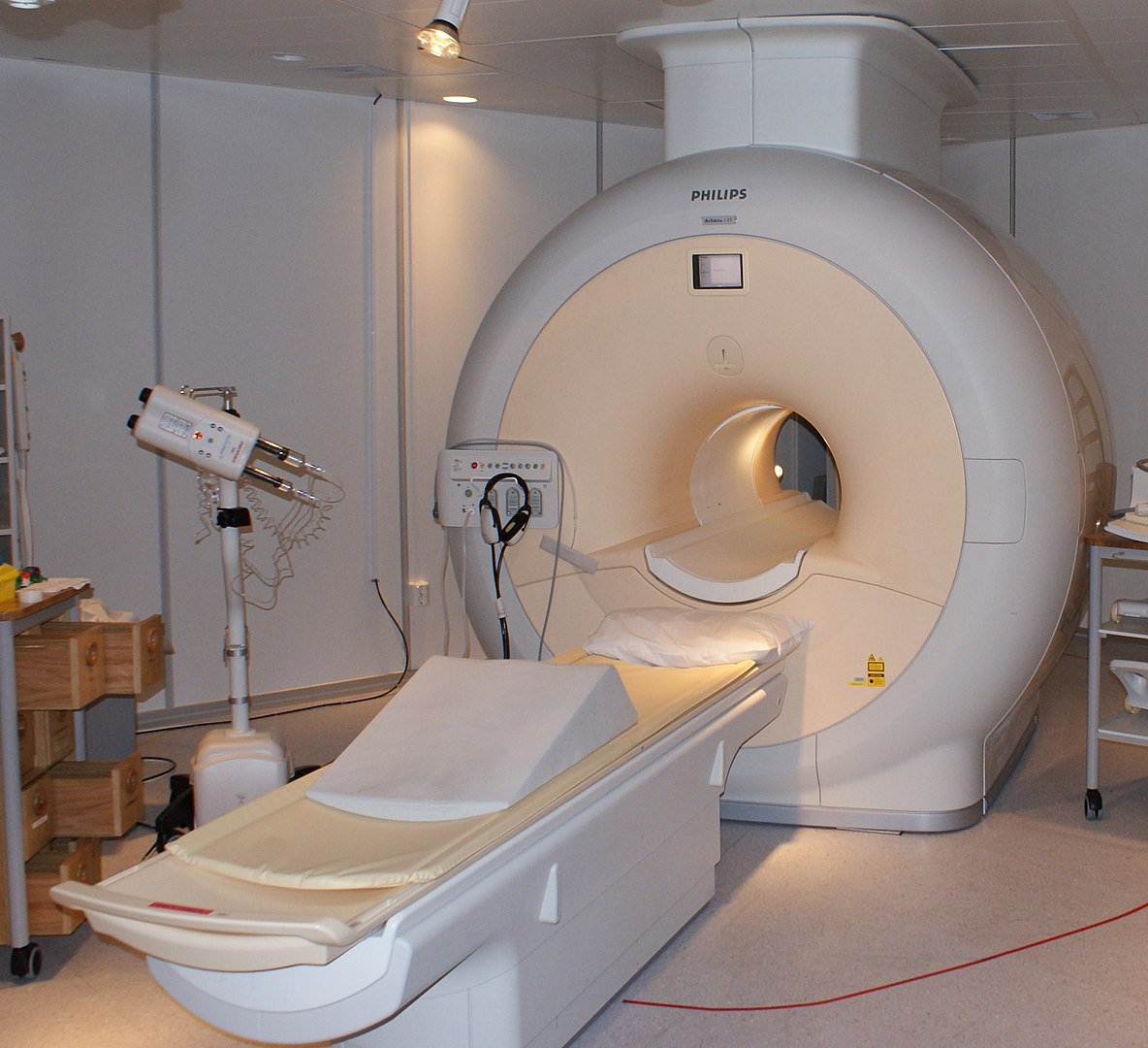
00:01 In this talk, we're going to discuss clinically isolated syndromes. 00:05 Those monophasic, CNS autoimmune conditions that occur. 00:11 Let's start with a case. 00:13 This a 51-year-old patient who developed left hand numbness and weakness progressing over the course of two weeks to the left leg. 00:21 She developed difficulty with walking and presented to the emergency department with gait dysfunction, left hemibody weakness, and mildly reduced light touch and vibration sensation. 00:33 Deep tendon reflexes are hyperreflexive in the left hemibody and normal and the right hemibody. 00:39 Mental status exam is normal. 00:41 Cranial nerve examination is also unremarkable. 00:45 So what do you think in this case? There's a few key features that should stick out in your mind. 00:51 The first is the onset of the symptoms. 00:53 This began over a two week interval. 00:55 That's a subacute onset condition. 00:58 And that's common for inflammatory disorders. 01:01 We can also see it with infections and occasionally some toxins and other conditions. 01:05 But in CNS inflammatory disorders should be one thing in our mind. 01:11 The second is gait dysfunction. 01:12 This patient has a left hemibody syndrome, sparing the cranial nerves and face as well as the brain. 01:19 And so this points us in the direction of spinal cord pathology. 01:24 And the last is the deep tendon reflexes which support that left hemibody syndrome and spinal cord localization. 01:32 So, what imaging would you perform to further evaluate this patient? MRI of the brain with and without contrast. 01:39 MRI of the cervical and thoracic spine with and without contrast. 01:43 EMG and nerve conduction study. 01:46 Or a CT of the head and treat the patient for an acute stroke. 01:51 Well, MRI of the brain may be done in this patient and it may be helpful, but without cranial nerve deficits or mental status symptoms, beginning with an MRI of the brain is probably not the best choice. 02:03 The patient's symptoms localize initially to the spinal cord and that's going to be the initial place we want to image. 02:10 CT of the head and treating this patient for a stroke would be a consideration for someone coming in with left hemibody symptoms. 02:17 But the timeline of onset is inconsistent with a stroke for this patient. 02:21 Strokes begin acutely over hours to maybe a day and this patient's symptoms developed slowly and progressively over two weeks and that's inconsistent with a vascular etiology. 02:33 EMG nerve conduction study can be very helpful in evaluating limb symptoms. 02:37 This patient has hyperreflexia, suggestive of a central nervous system disorder. 02:41 EMG and nerve conduction are typically normal with central nervous system disorders and are unlikely to reveal the causative pathology for this patient. 02:50 So an MRI of the cervical and thoracic spine is really the best next exam. 02:55 MRI of the brain is likely to also be performed but to figure out what's going on the MRI of the spine is where the money is. 03:03 And for this patient, they underwent MRI of the cervical and thoracic spine and here we're looking at a sagittal and an axial view of the MRI of the C spine. 03:14 In the sagittal sequences, on the left you can see a area of edema. 03:20 A new white matter lesion within the cord. 03:23 And on the axials, we see that it's eccentrically located on the left side of the cervical spine and causing this patient's left hemibody symptoms. 03:33 This imaging finding is suggestive of an inflammatory lesion but could also be seen with certains infections, or neoplasms, which would be on the differential for this patient. 03:45 Patient also underwent MRI of the brain to look for any other lesions that may be asymptomatic, new or old in this patient. 03:52 And here we're looking at representative cuts of the coronal FLAIR the fluid-attenuated inversion recovery image and an axial T2, which are essentially normal. 04:02 We don't see other white matter lesions that would be suggestive of prior insults, prior immune attacks on the brain in this patient. 04:10 And this is an important feature and aspect of the workup of a clinically isolated syndrome. 04:16 Clinically isolated syndromes, we see one attack. 04:19 And there's not evidence of prior insults in the brain or spine as we see here.
About the Lecture
The lecture Case: 51-year-old with Left Hand Numbness by Roy Strowd, MD is from the course Multiple Sclerosis (MS) and Inflammatory Disorders of the CNS.
Included Quiz Questions
Which statement best describes a clinically isolated syndrome?
- A single episode that progresses over days to weeks
- A single episode that progresses over minutes to hours
- Multiple episodes that are relapsing and remitting with an onset of days
- Multiple episodes that progress over weeks to months
Which imaging modality is most likely to reveal the inflammation when working up a clinically isolated syndrome?
- MRI
- CT scan
- Plain film X-ray
- Ultrasonography
Author of lecture Case: 51-year-old with Left Hand Numbness

Roy Strowd, MD
Customer reviews
5,0 of 5 stars
| 5 Stars |
|
5 |
| 4 Stars |
|
0 |
| 3 Stars |
|
0 |
| 2 Stars |
|
0 |
| 1 Star |
|
0 |
