Playlist
Show Playlist
Hide Playlist
Cardiac Case: 72-year-old Man with Dyspnea at Rest and Inability to Lie Flat
-
Cardiac Case 72-year-old Man with Dyspnea at Rest and Inability to Lie Flat.pdf
-
Reference List Cardiology.pdf
-
Download Lecture Overview
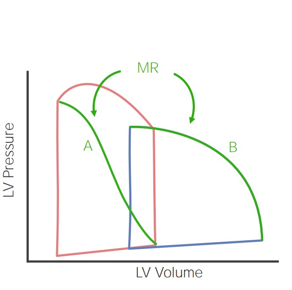
00:00 A 72-year-old man is admitted to the hospital because of dyspnea at rest and inability to lie flat due to shortness of breath. 00:08 Every time he tries to lie down he’s so short of breath he has to sit up. 00:11 He also has ankle swelling. 00:13 He reports these symptoms have been gradually developing over several months. 00:18 He lives with his wife and he's been well previously. 00:21 He was told as a young man that he had a heart murmur but it never caused him any trouble. 00:27 On physical exam his blood pressure was 139/75, not too remarkable for a man his age, heart rate is 88 and his fingertip oxygen saturation is a bit low at 88%. 00:39 His jugular venous pulse is elevated at 14 cm/h2o again implying a high pressure in the right atrium. 00:46 He has respiratory sounds, crackles 2/3 of the way up the back implying that there's a lot of fluid in the lungs and he has a grade 3/6 blowing holosystolic murmur best heard laterally in the apex and there's no third sound. 01:03 Let me imitate that for you. So here's normal, lub-dub, lub-dub, lub-dub. 01:09 Here it is with the murmur, lub-whoo-dub, lub-whoo-dub, lub-whoo-dub. 01:16 You can hear that sort of blowing murmur very different from the murmur of aortic stenosis which is hurm, hurm, hurm - this murmur is whoo, whoo, whoo; and is typical of the murmur of mitral regurgitation, there's no third heart sound and there's 2+ pitting edema consistent again with heart failure. 01:36 What are the critical findings here? Of course dyspnea at rest and inability to lie flat in other words orthopnea, are signs of heart failure. He's hypoxic which fits with his lung findings that is there's lots of fluid in the lung. 01:50 He has jugular venous distention, elevated right atrial pressure and of course he has the murmur of mitral regurgitation and his edema in his leg is another sign of heart failure. 02:02 So what are the diagnostic options? Well almost certainly one of the first things we do would be a Doppler and two-dimensional echo cardiographic study to see how good the ventricle is performing and to see if we could see the mitral regurge, and here we see two echos on the left is the image without the Doppler. 02:21 You can see the left ventricle is enlarged as is the left atrium and a little arrow points to the mitral valve that’s prolapsing and then in addition, you can see the Doppler collar lots of regurgitation into the left atrium so this is severe mitral regurgitation secondary to mitral valve prolapse. 02:40 So what's the options? Well, the first thing is because of his age, we do a catherization and we find that there is a atherosclerotic narrowing of the right coronary, severe mitral regurge and fortunately left ventricular function is normal. 02:55 so, the diagnosis is severe mitral regurgitation secondary to mitral valve prolapse and by ventricular heart failure. 03:03 Moving on to treatment, he's referred to cardio thoracic surgery where he undergoes a mitral valve repair and he gets a single coronary bypass to his right coronary. 03:13 One might have also just angioplasty then and stented the right coronary and then sent him just for the mitral valve repair and either one of those approaches would have been fine and the patient has an uneventful recovery and much less heart failure symptoms, is able to resume normal activities.
About the Lecture
The lecture Cardiac Case: 72-year-old Man with Dyspnea at Rest and Inability to Lie Flat by Joseph Alpert, MD is from the course Cardiovascular Cases.
Included Quiz Questions
Which of the following signs is associated with heart failure?
- Bilateral lower limb edema
- Caput medusa
- Rovsing's sign
- Cullen's sign
- Murphy's sign
Doppler echocardiography is most useful in the diagnosis of which condition?
- Valvular heart disease
- Pneumopericardium
- Coronary artery disease
- Arrhythmia
- Cardiac tamponade
Customer reviews
5,0 of 5 stars
| 5 Stars |
|
5 |
| 4 Stars |
|
0 |
| 3 Stars |
|
0 |
| 2 Stars |
|
0 |
| 1 Star |
|
0 |


