Playlist
Show Playlist
Hide Playlist
Cardiac Case: 62-year-old Man with Shortness of Breath and Peripheral Edema

-
Cardiac Case 62-year-old Man with Shortness of Breath and Peripheral Edema.pdf
-
Reference List Cardiology.pdf
-
Download Lecture Overview
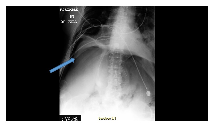
00:01 A 62-year-old man was admitted to the hospital because of marked shortness of breath and peripheral edema. 00:06 His personal history is that he’s had pneumonia a number of times over recent years, he smokes one to two packs of cigarettes a day for nearly 50 years and is still smoking and on physical exam he's thin, has a respiratory rate of 20 per minute which is borderline elevated. 00:24 He's hypertensive at 162/90 and his heart rate is somewhat increased at 92. 00:30 His peripheral oxygen saturation is poor at 84% on room air but on 4 L of oxygen its 91% so this is telling us already that there’s significant lung disease here. 00:43 His jugular venous pulse is also elevated at 14 so right atrial pressure is increased, he has distant long sounds and when you ask him to take a deep breath there’s not very good air movement. 00:56 His heart sounds are distant and hard to hear, he has an enlarged liver with the margin felt 4 cm below the right costal margin and he has 3+ to 4+ peripheral edema extending well up on the leg. 01:12 His EKG demonstrates right ventricular hypertrophy and a chest X-ray shows hyperinflated lungs with multiple areas of atelectasis or scarring presumably from a previous pneumonia. 01:24 So what are the critical things we want to think about here? First of all of course his short of breath and so that say heart or lung disease. 01:33 He's also had peripheral edema so we’re thinking possibly there’s gonna be heart failure involved here. 01:39 His history of all the smoking for many years tells us there’s liable to be lung possibly and heart disease. 01:46 He’s had pneumonia many times and of course he's still smoking and has a huge cigarette smoking history. 01:52 His respiratory rate is a little bit fast, his blood pressure is elevated, he's hypertensive and of course, he has markedly abnormal oxygen saturation suggesting that there is significant lung disease. 02:06 His JVP is elevated suggesting that there’s right ventricular failure with elevated right atrial pressure. 02:13 There are distant heart sounds with poor ventilatory capacity that’s telling us, okay we’ve got some science here that are gonna suggest very severe lung disease with some pulmonary hypertension possibly causing right-sided heart failure. 02:27 The heart sounds are also distant because there’s a lot of air trapping and again the enlarged liver suggests that’s congestive because of right ventricular failure and of course he also has edema. His EKG demonstrates right ventricular hypertrophy and this suggests that he has pulmonary hypertension leading to right ventricular failure. 02:48 His chest X-ray of course shows hyperinflated lungs with multiple areas of atelectasis or scarring - no surprise given his smoking history. 02:59 And here we see his EKG, you can see the very tall R wave in lead V1 suggesting pulmonary hypertension and that’s the cause of his peripheral edema and his right ventricular failure. 03:13 And you can see another characteristic there there’s deep S waves over the left precordial leads corresponding to the large R wave in lead V1. 03:25 There’s also right access deviation also part of the right ventricular hypertrophy EKG pattern and he has tall P waves so called P pulmonale commonly seen in advanced pulmonary disease, this is right atrial enlargement and remember jugular venous pulse told us - pressure told us that he had a high right atrial pressure so we’re not surprised to find right atrial enlargement on his EKG. 03:53 And the diagnoses are RVH, right ventricular hypertrophy and here we see a chest x-ray. 03:59 Notice how far the diaphragms are pushed down and notice how the heart is vertical - this is a man with sever emphysema. 04:08 The lung fields had minimal vascular markings, the lungs are hyperinflated and there the flatted diaphragms are typical chest x-ray from somebody with sever emphysema so chronic obstructive pulmonary disease and the echocardiogram shows a dilated right ventricle with decreased systolic function, left ventricle’s functioning fine. 04:31 The estimated pulmonary artery systolic pressure is 60 mmHg quite elevated so therefore and we know that’s what happens when people have severe end stage lung disease and of course his right atrial pressure is elevated. 04:46 So the diagnosis is right ventricular failure secondary to pulmonary hypertension which is the result of severe chronic obstructive pulmonary disease. 04:56 This is called cor pulmonale that is the lung heart and of course we know smoking causes damage to the lungs and eventually leads to pulmonary hypertension. 05:06 The left ventricle is normal unless there’s concomitant coronary disease which could happen in somebody he smokes this much and this form of heart failure as I mentioned is called cor pulmonale -- heart failure secondary to lung disease. 05:20 Treatment: diuretics help with getting the liver congestion down and the peripheral edema, we want to maximize lung function so he’s got to stop smoking. 05:30 Cor pulmonale doesn’t respond to the same medications used for systolic left ventricular failure so in the end diuresis and pulmonary rehab would be the best treatments for him.
About the Lecture
The lecture Cardiac Case: 62-year-old Man with Shortness of Breath and Peripheral Edema by Joseph Alpert, MD is from the course Cardiovascular Cases.
Included Quiz Questions
A 62-year-old man is admitted to the hospital because of marked shortness of breath and peripheral edema. The patient is a heavy smoker, and he has had pneumonia numerous times over many years. On physical examination, he appears thin, his respiratory rate is 20 beats/min, his blood pressure is 162/90 mm Hg, his heart rate is 92 beats/min, and his oxygen saturation 84% on room air but 91% on 4 liters of oxygen by nasal cannula. His jugular venous pulse is at 14 cm of water. On auscultation, distant lung sounds with poor ventilatory capacity and distant, hard-to-hear heart sounds are noted. On palpation, the liver is felt to be enlarged, with its margin 4 cm below the right costal margin. 3–4 peripheral edema extending to the thigh is noted. An ECG and chest x-ray are performed. Which of the following will most likely be found on ECG?
- Tall R waves in V1
- Deep S waves in V1 and V2
- Left-axis deviation
- Peaked T waves in V1–V6
- Diffuse elevation of the ST-segment
A 62-year-old man is admitted to the hospital because of marked shortness of breath and peripheral edema. The patient is a heavy smoker, and he has had pneumonia numerous times over many years. On physical examination, he appears thin, his respiratory rate is 20 beats/min, his blood pressure is 162/90 mm Hg, his heart rate is 92 beats/min, and his oxygen saturation 84% on room air but 91% on 4 liters of oxygen delivery by nasal cannula. His jugular venous pulse is at 14 cm of water. On auscultation, distant lung sounds with poor ventilatory capacity and distant, hard-to-hear heart sounds are noted. On palpation, the liver is felt to be enlarged, with its margin 4 cm below the right costal margin. 3–4 peripheral edema extending to the thigh is noted. An ECG shows right ventricular hypertrophy, and a chest x-ray indicates hyperinflated lungs with multiple areas of atelectasis or scarring. Which of the following is the most likely diagnosis?
- Cor pulmonale
- Systolic left heart failure
- Constrictive pericarditis
- Liver failure
- Pulmonary fibrosis
A 62-year-old man is admitted to the hospital because of marked shortness of breath and peripheral edema. The patient is a heavy smoker, and he has had pneumonia numerous times over many years. On physical examination, he appears thin, his respiratory rate is 20 beats/min, his blood pressure is 162/90 mm Hg, his heart rate is 92 beats/min, and his oxygen saturation 84% on room air but 91% on 4 liters of oxygen delivery by nasal cannula. His jugular venous pulse is at 14 cm of water. On auscultation, distant lung sounds with poor ventilatory capacity and distant, hard-to-hear heart sounds are noted. On palpation, the liver is felt to be enlarged, with its margin 4 cm below the right costal margin. 3–4 peripheral edema extending to the thigh is noted. An ECG shows right ventricular hypertrophy, and a chest x-ray indicates hyperinflated lungs with multiple areas of atelectasis or scarring. After an echocardiogram, the diagnosis of cor pulmonale is established. Which of the following will most likely be seen on an echocardiogram?
- Elevated right atrial pressure
- Decreased left ventricular systolic function
- Decreased pulmonary artery systolic pressure
- Right ventricular collapse
- Dilated left atrium
Author of lecture Cardiac Case: 62-year-old Man with Shortness of Breath and Peripheral Edema

Joseph Alpert, MD
Customer reviews
5,0 of 5 stars
| 5 Stars |
|
5 |
| 4 Stars |
|
0 |
| 3 Stars |
|
0 |
| 2 Stars |
|
0 |
| 1 Star |
|
0 |
