Playlist
Show Playlist
Hide Playlist
Breast Cancer: Mammographic Signs

-
Slides Breast Female Repro.pdf
-
Download Lecture Overview
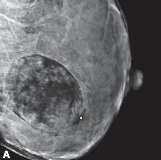
00:01 In this section, I want to quickly go over every important screening test. 00:07 Remember please that at some point in time, unfortunately, if a female goes onto develop breast cancer, incidence-wise, remember it’s number one. 00:15 Mortality-wise, it’s number two. 00:17 So even with proper amounts of screening and even with proper medications, it’s still the number two killer of cancer in a female. 00:28 Let’s do a mammography. 00:30 You’re looking for a mass, you’re looking for architectural distortion. 00:35 And you’re looking for calcification. 00:37 What you’re looking at here in your mammography would be one in which – Well, you’ll notice that the anterior portion will be nipple. 00:45 And the one on the right will be normal. 00:48 What may then happen is that if breast cancer was to kick in or was to develop and at some point in time, maybe there might be retraction. 00:56 Oftentimes, that structure of the breast that is retracted on your very right. 01:01 Now, are you picturing this? Are you able to close your eyes and conceptualize what this looks like? The anterior portion would be on the very right and as you move from right to left, you’re moving from the nipple. 01:11 And then in a little bit, I’ll walk you through the pathophysiology or really the clinical anatomy of the breast apparatus. 01:19 And as you move from the nipple, you’re coming into lactiferous sinus, then you’re coming into your major duct, terminal duct and then you go into lobules. 01:26 And then in the very back on your left will be the stroma that’s responsible for holding onto the breast or you have these Cooper's ligaments. 01:34 Remember those in anatomy? The suspensory ligaments that hold on to the breast. 01:39 And when a female gets old enough, then the suspensory ligament becomes weaker and then literally causes ptosis of the breast or sagging breasts. 01:47 You call that a Cooper’s breast. 01:49 Now that you understand the picture of what you’re looking at here on mammography, if there’s retraction of the nipple then on the left it’s kind of showing you that with increased fibrosis. 01:59 In addition, there might be calcification in which the calcium is then coming through the ducts. 02:04 Let’s spend more time here and just because you see calcification though on mammography, which appears as being white, right? It’s always white, the calcium is. 02:13 And it doesn’t always mean breast cancer though and that’s where the boards will trick you. 02:17 But they won’t though, they won’t trick you because you’ll know better.
About the Lecture
The lecture Breast Cancer: Mammographic Signs by Carlo Raj, MD is from the course Reproductive Pathology: Breast Disease with Carlo Raj.
Included Quiz Questions
Which of the following statements about mammographic signs is CORRECT?
- Architectural distortions may be a sign of cancer.
- Calcium deposits are radiolucent.
- Calcifications on mammography are always indicative of breast cancer.
- Weakening of Cooper's ligament is visible by mammography.
- Masses seen on mammography are always malignant.
Author of lecture Breast Cancer: Mammographic Signs

Carlo Raj, MD
Customer reviews
2,8 of 5 stars
| 5 Stars |
|
1 |
| 4 Stars |
|
0 |
| 3 Stars |
|
1 |
| 2 Stars |
|
1 |
| 1 Star |
|
1 |
Very Nice Lecture. He has a unique way of teaching topics and make us remember stuff and give a foundation on medicine. His Integrative Teaching method is very Good. Perfect Lecturer for Pathology
Ok explanation. Some examples and pictures to explain the topic would make it better
not good at all poor presentation lack of information sorry but this is the truth
Average, alright, acceptable, fair, reasonable, of an acceptable standard, overall underwhelming
