Playlist
Show Playlist
Hide Playlist
Basal Cell Carcinoma: Diagnosis and Management

-
Reference List Pathology.pdf
-
Slides Basal Cell Carcinoma Diagnosis Management.pdf
-
Download Lecture Overview
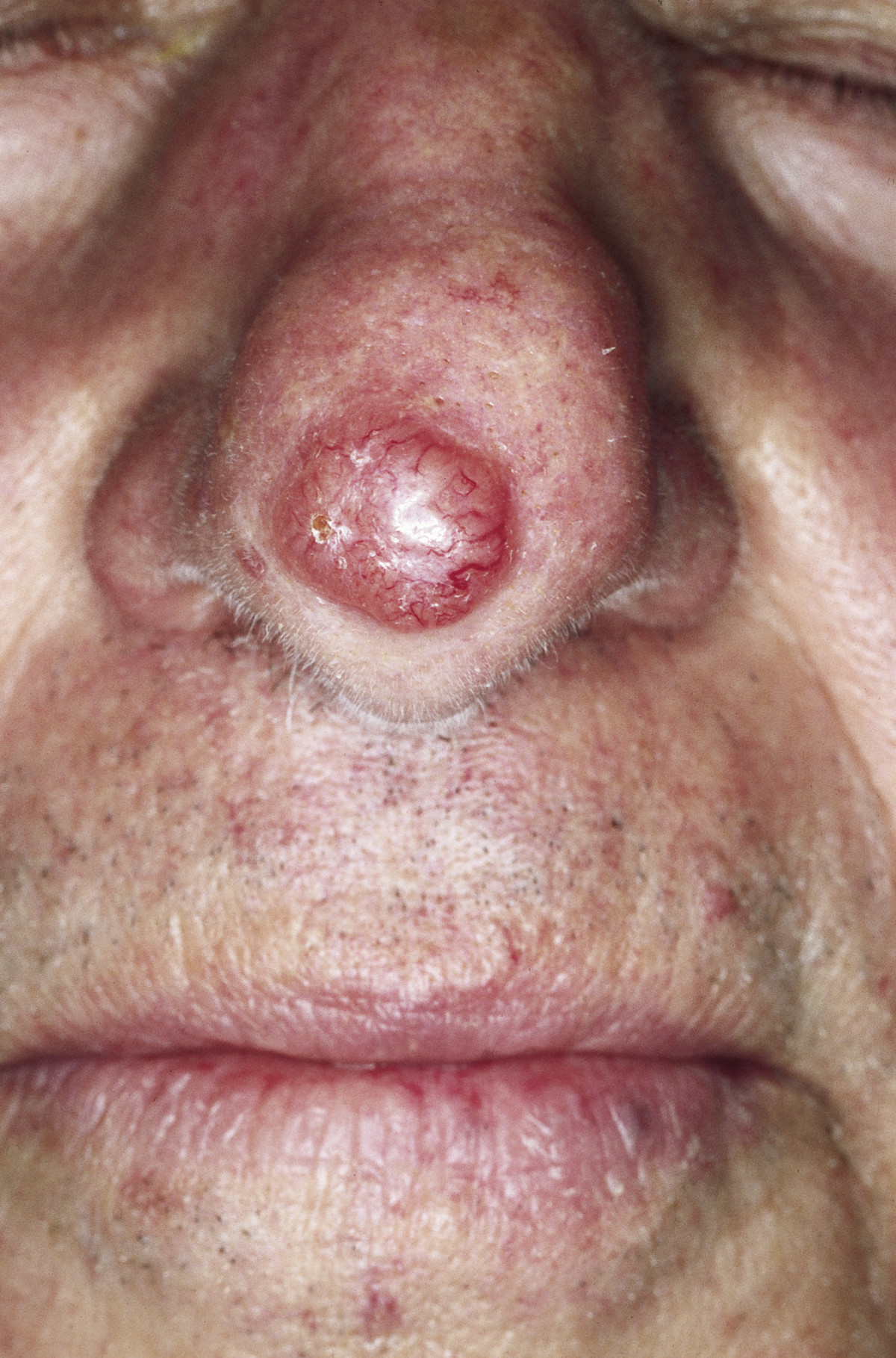
00:01 So clinical presentation. 00:04 The disease characteristics of basal cell carcinomas, they can look like a lot of different things. 00:10 They are the classical versions that I'm going to show you. 00:13 But there are some kind of atypical ones. 00:15 So you have to have a reasonably high index of suspicion in order to do a biopsy and diagnose it. 00:22 They can be small. 00:24 They can be larger. 00:25 They are typically slow growing. 00:28 Most remain localized. 00:30 But even in that localized place, they can be very aggressive locally. 00:35 They tend not to metastasize. 00:38 So those locally aggressive ones, you're seeing a good example there adjacent to this guy's earlobe. 00:45 That in fact is what we describe as a rodent ulcer. 00:48 Why is it called that? Well, somebody in the past decided that's what your skin would look like if a rodent chewed on it. 00:57 Whatever. 00:58 Anyway, that can be locally invasive and especially in places on the skin around the eyes, the nose, etc. 01:04 can be quite disfiguring. 01:06 As I already said, it's exceptional. 01:10 It's reportable if a basal cell carcinoma metastasizes. 01:15 And overall, it has a very, very low mortality, again, associated with those tumors that do metastasize. 01:23 It is locally aggressive. 01:25 It is malignant, but it doesn't metastasize very much. 01:29 It can look in various ways that we're going to see on the following slides. 01:34 The face, because of all the sun exposure the face gets, is a typical location for it. 01:40 And it's usually over the nose, the bridge of the nose, the male or eminences, the forehead, nasolabial folds, all things that are getting sun exposure. 01:50 Trunk and the other remaining body parts also clearly, depending on sun exposure, as a major driving force, can develop basal cell carcinomas. 02:00 The histopathology, so remember it's basal cells. 02:04 So the tumors look like the basal cells at the derma-epidermal junction. 02:09 It's a proliferation of those. 02:12 They tend to kind of pile up, as you see here. 02:16 And they can have also, because the cells are a little bit weird and may not be fully survivable in terms of their mutations, you may see cellular apoptosis. 02:30 The histologic types, so you can have nodular ones that kind of stick up. 02:35 You see that on the end of the guy's nose. 02:37 You can have kind of superficial that look fairly flat. 02:43 You can have morphia form. 02:45 That means that they're kind of fibrosine or sclerosine. 02:48 And other rarer types are described there, basal squamous and pigmented basal cell carcinomas. 02:55 Those are all variants. 02:56 The most typical type is that nodular or superficial basal. 03:01 The diagnosis, so if there's a family history, yes. 03:05 So that may actually speak to whether or not you have germline patch mutations. 03:10 But if you've had previous skin cancers or you've been in the sun, yeah, it's very likely that that new thing that's growing is a basal cell carcinoma. 03:21 Individuals who are HIV positive or otherwise have an immunosuppressed status, say due to chemotherapy, are also prone to basal cells because we don't have the normal immunologic mechanisms by which we can surveil and destroy them. 03:38 Physical exam, you need to look at the entire skin top to bottom. 03:42 And you may want to assess for palpable lymphadenopathy. 03:46 Again, really rare that these ever metastasize. 03:51 Dermoscopy can actually help. 03:53 You can identify vessels. 03:55 So there's vasculogenesis that occurs with these. 03:59 You can see evidence of kind of nests of cells deep to the superficial dermis. 04:05 You can see ulceration and lack of pigmented network. 04:11 So if there's pigmented network, you tend to think more of a melanocytic lesion, melanoma. 04:18 But the lack thereof may help you in making a diagnosis of a basal cell. 04:25 Ultimately, it comes down to let's do a biopsy. 04:28 Let's send things off to the pathologist. 04:30 And you can do it either as a shave, a punch, or a completely excisional biopsy. 04:35 How do we manage these? Well, so again, because although they can be locally aggressive, they don't metastasize, standard surgical excision is usually curative. 04:47 You want to get larger margins so that you don't have it recurring in the original site. 04:52 But usually, that is more than enough. 04:55 For areas where it is high risk because of certain features in terms of vascularity and things like that, you may want to do what's called a Mohs micrographic surgery. 05:08 And Mohs involves taking a little shave, send it to pathology and say, "Is there tumor or is there not tumor?" And then they say, "There's tumor." And you take another shave. 05:17 You take another shave. 05:18 And eventually, after multiple kind of going back and forth with pathology, say, "There's no more tumor. You got it all." That's important for things that are potentially high risk, but also is done for facial lesions where you don't want to take out big hunks of skin, say, in the middle of the face adjacent to the nose or to the eye. 05:37 So you'll do Mohs surgery for that. 05:40 Alternative therapies, you can do chemotherapy with a variety of agents. 05:44 You can do electrodesiccation and curatage. 05:47 You can put liquid nitrogen on it. 05:49 You can radiate them. 05:51 But usually, unless it's in a very difficult to get at area, say, on the face, you would just do an excisional treatment. 06:00 And with that, we've covered a really common entity, a very important malignant entity, and one you will undoubtedly encounter in your lifetime, basal cell carcinomas.
About the Lecture
The lecture Basal Cell Carcinoma: Diagnosis and Management by Richard Mitchell, MD, PhD is from the course Premalignant and Malignant Epidermal and Dermal Tumors.
Included Quiz Questions
Which of the following best describes the typical growth pattern of basal cell carcinoma?
- Slow-growing and locally aggressive with rare metastasis
- Rapidly growing with frequent metastasis
- Slow-growing with frequent metastasis
- Rapidly growing but always remains superficial
- Self-limiting with spontaneous regression
Which facial location is most commonly affected by basal cell carcinoma?
- Bridge of the nose
- Upper eyelid
- Lower lip
- Behind the ear
- Chin
When would Mohs micrographic surgery be most appropriate for treating basal cell carcinoma?
- For facial lesions where tissue preservation is crucial
- For small lesions on the trunk
- For metastatic disease
- For multiple superficial lesions
- For lesions on the extremities
Author of lecture Basal Cell Carcinoma: Diagnosis and Management

Richard Mitchell, MD, PhD
Customer reviews
5,0 of 5 stars
| 5 Stars |
|
5 |
| 4 Stars |
|
0 |
| 3 Stars |
|
0 |
| 2 Stars |
|
0 |
| 1 Star |
|
0 |
