Playlist
Show Playlist
Hide Playlist
Auditory Nuclei, Auditory Pathway and Otosclerosis

-
Slides 9 AuditorSystem BrainAndNervousSystem.pdf
-
Reference List Anatomy.pdf
-
Download Lecture Overview
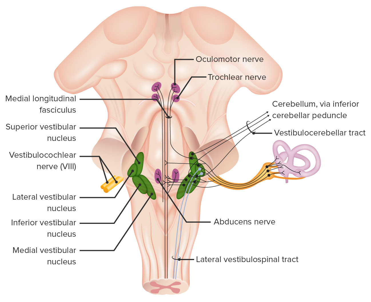
00:00 The afferent fibers conveying information from the organ of Corti are going to convey this information to auditory nuclei. These will be the conductors of beautiful music or audition. There are several auditory nuclei that are conductors of audition. First, we have up here superiorly, the medial geniculate body. 00:30 This is a thalamic nucleus. Another member of the auditory nuclei is the inferior colliculus that we see in through here. 00:42 Third member is the nuclei that are associated with the lateral lemniscus. We also have cochlear nuclei in this area. Then we have the olivary nuclei. These are paired structures hence, plural. Now, I want you to understand the circuitry of the auditory pathway. First again will be depolarization of the cochlear hair cells in the cochlea. Action potentials will then be conveyed along the cochlear nerve fibers to the ipsilateral anterior cochlear nucleus. That has been highlighted here. From this nucleus, fibers then will travel to the superior olivary nucleus on the same and opposite sides. Here is the superior olivary nucleus on the same side receiving information from the auditory nucleus. Then there’s also signaling taking place to the superior olivary nucleus on the opposite side as well. Synapses occur within here. 02:10 Then neurons will then travel through the lateral lemniscus from the cochlear and superior olivary nuclei. 02:24 Their destination is to the inferior colliculus that we see here. Then from the inferior colliculus, there will be another neuron that will convey auditory information to the medial geniculate body of the thalamus. Then synapses within this body will send auditory signals to the primary auditory cortex which is associated with the transverse temporal gyrus that’s a part of the temporal lobe of the cerebrum. There can be some lesions associated along the auditory pathway that can impair hearing. 03:15 So, it’s important for you to understand those in a clinical context. The first area that’s going to be lesioned is going to be that at the green line. So here, we’re having a lesion along the cochlear or the vestibular cochlear component of cranial nerve number eight. If this is a unilateral lesion, an individual will have sensorineural hearing loss on that side. Lesions either here at the black line or even more superiorly here on the contralateral side will result in bilateral sensorineural diminishment of hearing. Individuals that have lesions along these pathways at the black lines are also going to have a poor ability to localize the source of the sound. Now, we have an opportunity for you to understand some clinical considerations. Let’s talk about for a moment about conductive hearing loss. Conductive hearing loss is essentially poor transmission from the external to the middle ear. There are several causes for this poor transmission. 04:40 One may be the fusion of the ear ossicles together which decreases their ability to conduct sound from the tympanic membrane to the oval window. This, in and of itself, is called otosclerosis. 04:58 If there’s impacted wax in the external acoustic meatus, that may make it more difficult for sound to strike the tympanic membrane resulting in poor transmission. If the tympanic membrane is perforated, this too will lead to a poor transmission. Then fluid that accumulates in the middle ear makes it much more difficult as well for there to be transmission from the external to the middle ear.
About the Lecture
The lecture Auditory Nuclei, Auditory Pathway and Otosclerosis by Craig Canby, PhD is from the course Auditory System and Vestibular System. It contains the following chapters:
- Auditory Nuclei
- Auditory Pathway
- Otosclerosis
Included Quiz Questions
To which of the following do the cochlear nerve fibers transmit nerve impulses?
- Ipsilateral anterior cochlear nucleus
- Contralateral superior olivary nucleus
- Medial geniculate body
- Superior olivary nucleus
- Lateral geniculate body
In which of the following is the primary auditory cortex located?
- Temporal lobe
- Parietal lobe
- Postcentral gyrus
- Occipital lobe
- Central sulcus
Which of the following would be caused by a lesion of the inferior colliculus (central lesion)?
- Inability to locate the source of sound
- Inability to hear high-frequency sounds only
- Inability to hear low-frequency sounds only
- Conductive hearing loss on the ipsilateral side
- Unilateral sensorineural hearing loss
Which of the following characterizes otosclerosis?
- The fusion of middle-ear ossicles
- Fluid accumulation in the middle ear
- Impacted wax
- The fusion of hair cells
- Fibrosis of hair cells
Author of lecture Auditory Nuclei, Auditory Pathway and Otosclerosis

Craig Canby, PhD
Customer reviews
5,0 of 5 stars
| 5 Stars |
|
1 |
| 4 Stars |
|
0 |
| 3 Stars |
|
0 |
| 2 Stars |
|
0 |
| 1 Star |
|
0 |
Straightforward, explanative, and helpful It would be enhanced with the inclusion of labeled real anatomical structures to supplement learning and retention.
