Playlist
Show Playlist
Hide Playlist
Aortic Arches: Morphologic Changes

-
Slides 06-32 Aortic Arches and Large Arteries.pdf
-
Reference List Embryology.pdf
-
Download Lecture Overview
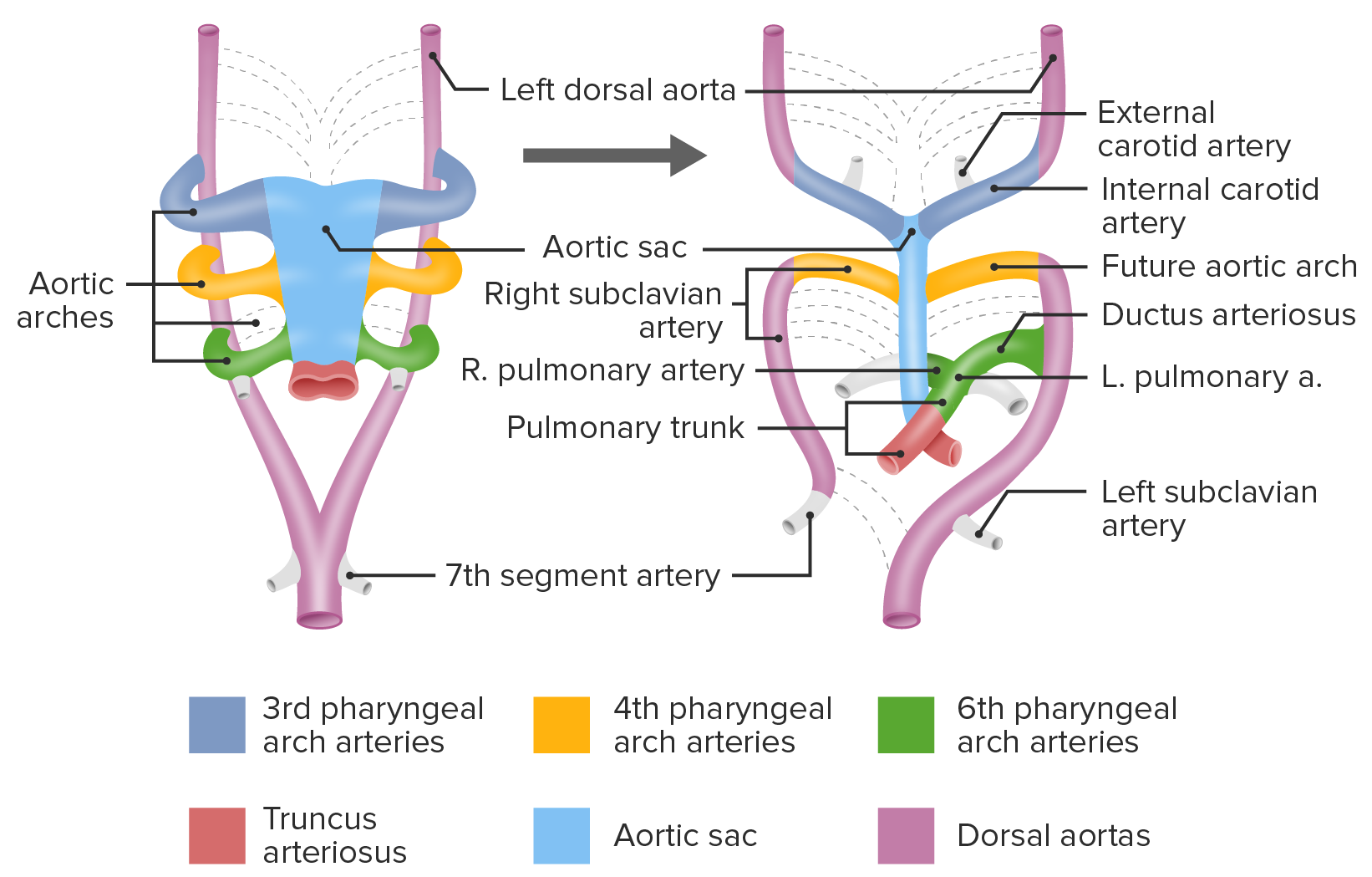
00:01 So let's return to this image and put that schematic to work. 00:04 We can see that right here the aorta is gonna propel blood up to 3rd arch to the head, 4th arch to the right upper limb and the left side of aortic arch and then here, when the pulmonary trunk receives blood from the right ventricle it's gonna push into the two pulmonary arteries but potentially also through the ductus arteriosus into the aorta. 00:30 On the left side, the 6th arch loses its connection to the dorsal aorta and the 4th arch also retains its connection above there. 00:41 However, a small portion of the dorsal aorta on the right is going to die off and that's going to then detach from the rest of the aorta. 00:53 On the right side, something called the 7th intersegmental artery is the last place we have that dorsal aortic connection and that 7th intersegmental artery is going to become the subclavian artery or the main blood supply to the upper limb. 01:07 So in this picture it's easy to think of these vessels just floating free in space, but I want you to remember that the 7th intersegmental artery on both the right and left is going to the upper limb and it's tethered in space where it can no longer move any further out and it's not gonna be able to just move around freely. 01:24 So here, when we move to the mature circulation we've got the aorta and pulmonary trunk leaving the heart. 01:33 Then the aortic arch, brachiocephalic trunk are both derived from aortic sac, at least very proximally. 01:40 The common and parts of the internal and external carotid arteries are then be coming from the 3rd aortic arch. 01:46 On the right side, the 4th aortic arch is going to contribute to the subclavian artery, but the rest of the subclavian arteries coming from the right 7th intersegmental artery. 01:57 Now we move a little bit further along the aortic arch. 02:00 It's gonna be derived partially from the 4th aortic arch on the left and the segment of the dorsal aorta shown here in purple that was present all along on the posterior side of the body wall. 02:14 The left subclavian artery is gonna be derived from the 7th intersegmental artery on the left and its direct connection to the dorsal aorta. 02:23 And then the right and left pulmonary arteries are both coming from the 6th arch, as is that connection between the pulmonary trunk and the aorta, only on the left the ductus arteriosus. 02:37 So let's take a quick look at all of that. 02:39 I know the terminology is confusing so let's just go back to the image and I'll narrow it along with what's happening. 02:45 We've got the 3rd, 4th and 6th aortic arches leaving the aortic sac and travelling to a right and left dorsal aorta. 02:53 The arrows indicate a place where there's gonna be a pinch and the vessels are going to narrow and eventually detach, so the dorsal aorta between the 3rd and 4th arches on both sides will narrow. 03:05 And we move to the picture on the right and you can see that the 3rd and 4th arches are moving apart. 03:10 The 3rd arch could become the common carotid arteries. 03:13 Next thing is we're gonna have narrowing and eventual loss of the connection of the 6th arch on the right to the dorsal aorta and the dorsal aorta is going to rescind between the upper limb, 7th intersegmental artery on the right and the fused dorsal aorta. 03:32 So we move one step further and we see this, and it's starting to look a bit like a mature circulation. 03:38 Recall that the 6th aortic arch is gonna be providing blood to the lungs, so that's now illustrated as well. 03:45 The 6th arch has no connection with the dorsal aorta on the right but it retains its connection on the left and that's gonna become the ductus arteriosus. 03:54 And as the body lengthens and those rescinded areas of the artery just come free, we move to the image on the right and we're left with mature circulation. 04:03 And just to run through it one more time, 3rrd arch is going to be right and left common carotid arteries and some portions of the internal and external thereafter. 04:13 4th arch on the right contributes to the subclavian artery. 04:16 4th arch on the left contributes to the arch of the aorta. 04:19 The two distal subclavian arteries on both right and left are coming from the 7th intersegmental artery that was supplying the body anyway. 04:29 And lastly, the 6th aortic arch is gonna contribute the pulmonary arteries on both right and left and only on the left side that ductus arteriosus that connects the pulmonary trunk to the aorta. 04:42 That's gonna be incredibly important when we talk about the transition from fetal circulation into adult circulation.
About the Lecture
The lecture Aortic Arches: Morphologic Changes by Peter Ward, PhD is from the course Development of Thoracic Region and Vasculature.
Included Quiz Questions
From what structure does the left subclavian artery derive?
- Left 7th intersegmental artery
- 6th pharyngeal arch
- Left dorsal aorta
- Left 4th aortic arch
- Left 3rd pharyngeal arch
From what structure do the aortic arch and brachiocephalic trunk derive?
- Aortic sac
- 1st aortic arch
- 2nd aortic arch
- 3rd aortic arch
- 6th aortic arch
The lungs will receive blood from the arteries that are derived from which arch?
- 6th arch
- 1st arch
- 2nd arch
- 3rd arch
- 4th arch
Author of lecture Aortic Arches: Morphologic Changes

Peter Ward, PhD
Customer reviews
5,0 of 5 stars
| 5 Stars |
|
2 |
| 4 Stars |
|
0 |
| 3 Stars |
|
0 |
| 2 Stars |
|
0 |
| 1 Star |
|
0 |
This was a great lecture! The only thing I missed was an overview in the form of a table or something to help with the review of what aortic arches gives rise to which arteries.
Your images really helped me visualize this process. Thank you
