Playlist
Show Playlist
Hide Playlist
Ankle Joint – Joints of Lower Limb

-
Slides 09 LowerLimbAnatomy Pickering.pdf
-
Download Lecture Overview
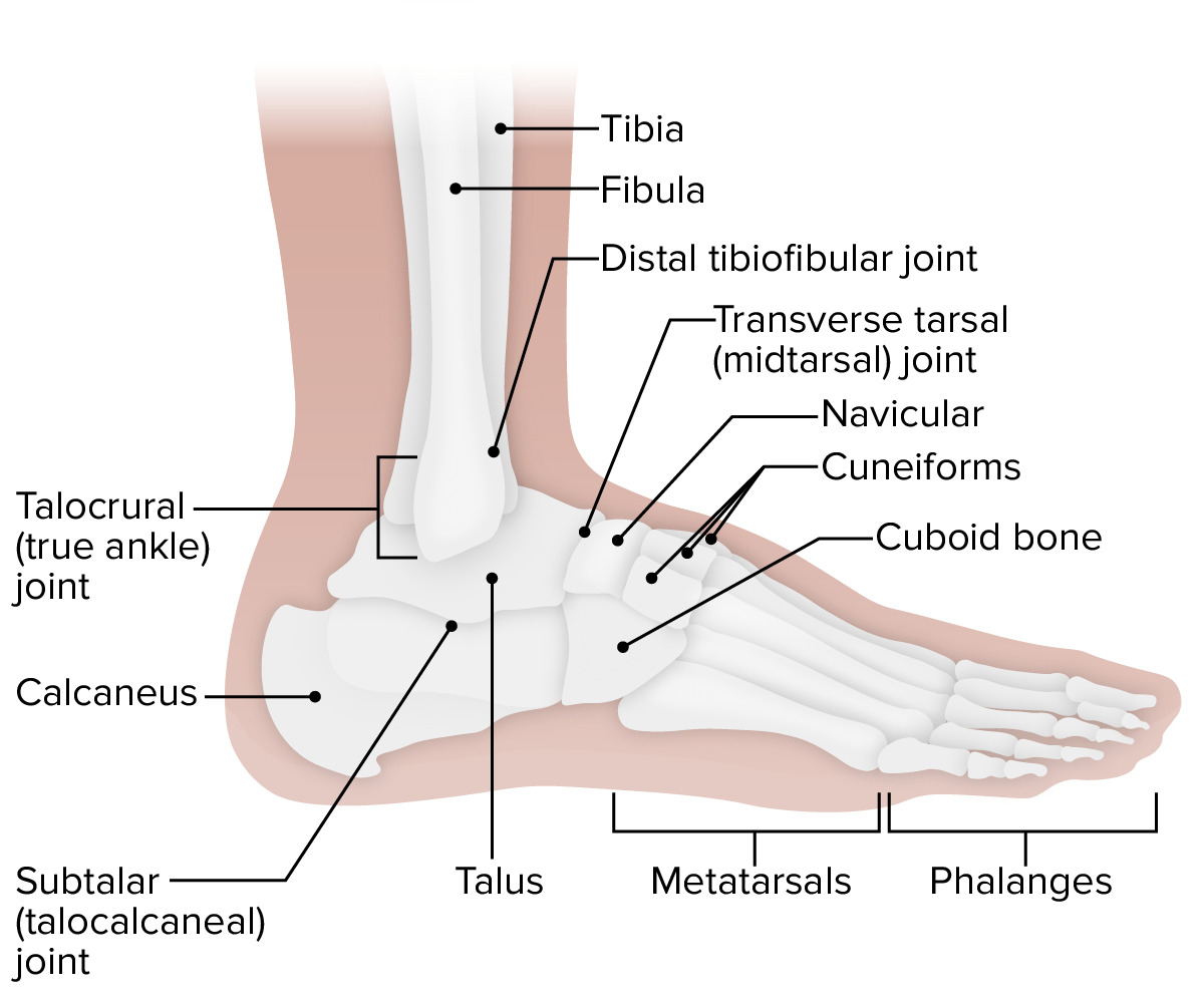
00:01 So here we can see, as we go to the ankle joint, this anterior tibiofibular ligament, and here we can see the posterior tibiofibular ligament. And here we can see the two articular facets of the lateral malleolus and the medial malleolus here. 00:17 So this is where we’re going to have the articulation with the trochlear surface on the talus, and we can see that here. So the joint capsule for the ankle joint is relatively thin anteriorly and posteriorly enabling dorsiflexion and plantarflexion to occur. There are heavy supports lateral and medially via collateral ligaments as we’ll see. Superiorly, the joint capsule attaches to the articular margins of the tibia and fibula. And inferiorly, they attach to the talus away from those articular surfaces. The articulations at the ankle joint are the distal tibia and fibula with the trochlea of the talus. So here we can see the talus, and here we have this smooth trochlear surface. And the distal end of the tibia and fibula form a deep socket, and this is known as the malleolar mortise. So with the medial malleolus and the lateral malleolus bulging away from the distal ends of the tibia and the fibula, they form this mortise, and that sits on the rounded superior surface of the talus. 01:32 Here, we can see the fibula medial surface of the lateral malleolus with the lateral talus. 01:40 So here, we can see this medial surface of the lateral malleolus is going to articulate with the lateral surface of the talus. The roof of the malleolar mortise is going to go to the talus. So here we see the roof of the malleolar mortise, and that’s articulating directly on top of the talus. And the medial malleolus, which we can see here, is going to articulate with the medial talus. During dorsiflexion, the joint is more stable as the anterior talus is wider driving into the tibia and fibula. Here, if you see, the anterior talus is wider and it tapers away posteriorly into this thinner region. With dorsiflexion, the malleolar mortise is going to be sitting against this wider anterior portion, and this wedges into this malleolar mortise, with the anterior and posterior tibiofibular ligaments holding the joint tightly in position. So with dorsiflexion, the joint becomes a lot more stable due to the wider anterior aspect of the talus. If we carry on looking at the ankle joint, we can see that it’s supported by a number of ligaments. Here again, we can see the posterior talofibular ligament, and we can also see we have a series of medial and lateral deltoid ligaments. So if we look at the ligaments of the ankle joint, the lateral ligament is formed by three separate ligaments. We have the anterior talofibular. Here, we can see the anterior talofibular. This is running from the lateral malleolus to the neck of the talus, and it’s weak, a flat band. We also have the posterior talofibular ligament. We can see it here. And this is running from the malleolus of the fibula to the lateral tubercle of the talus. It is a thick strong band. It is the posterior talofibular. 03:52 We can also see the calcaneofibular. Here, we can see the calcaneofibular ligament. 04:00 And that’s running from the lateral malleolus, the distal end of the fibula to the lateral aspect of the calcaneus. And here we have a rounded cord. So the lateral ligaments, we’ve got these three separate ligaments: anterior talofibular, posterior talofibular, and calcaneofibular. If we look at the medial or deltoid ligament, then this is a large strong ligament that originates from the medial malleolus part of the tibia. It spreads out distally to the talus, the calcaneus, the navicular, and it serves to stabilize the ankle joint during eversion. Remember, this was when your foot is lifted so that your little toe is lifting upwards and your big toe is flat on the floor. We have a few ligaments that make up this medial one. We have the tibionavicular ligament. So here, we can see the tibionavicular ligament just running down here. It’s very small, running from the tibia to the navicular bone. Here, we can see the tibiocalcaneal ligament. 05:11 So it’s running down from the tibia to the calcaneus. Between them, we can see the anterior tibiotalar ligament running from the tibia to the talus. And most posteriorly, we can see the posterior tibiotalar ligament. So we have these ligaments that are reinforcing the medial aspects of the joint. And this is important during eversion. Remember, when your little toe is lifted up and the medial aspect of your ankle is stretched, and this can help to control that aspect of the ankle joint.
About the Lecture
The lecture Ankle Joint – Joints of Lower Limb by James Pickering, PhD is from the course Lower Limb Anatomy [Archive].
Included Quiz Questions
Plantar flexion and dorsiflexion are aided by which portions of the thinned joint capsule?
- Anterior and posterior
- Medial and lateral
- Superior and inferior
- Superficial and deep
- Anterior and medial
Which surface connects to the inferior attachment of the ankle joint capsule?
- Talus
- Articular margin of tibia
- Articular margin of fibula
- Malleolar mortise
- Trochlea
Which feature gives stability to the ankle joint in dorsiflexion?
- Wider aspect of the talus
- Narrower aspect of the talus
- Tight holding of the posterior tibiofibular ligament
- Flexibility in the tibiofibular ligament
- Short length of the tibiofibular ligament
Author of lecture Ankle Joint – Joints of Lower Limb

James Pickering, PhD
Customer reviews
1,0 of 5 stars
| 5 Stars |
|
0 |
| 4 Stars |
|
0 |
| 3 Stars |
|
0 |
| 2 Stars |
|
0 |
| 1 Star |
|
1 |
terrible again... not even written what kind of joint it is!!
