Playlist
Show Playlist
Hide Playlist
Anatomy of the Teeth

-
Slides Head Neck Anatomy Teeth.pdf
-
Download Lecture Overview
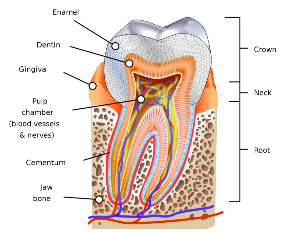
00:00 This presentation is on the oral cavity. Our specific focus will be on the teeth. The first up here is to look at the types of teeth that we have in the upper and lower jaws. Here, we're seeing a view of the upper or maxillary jaw. Each jaw, maxillary or upper has 16 teeth and then the lower jaw or mandibular jaw will also have 16 teeth. Of the 16 teeth, 4 of them are going to be incisors on the upper and the lower jaw. So here we have the 4 incisors located anteriorly and starting to spread laterally. Next, we have a pair of canines. 00:49 Here is the one on one side of the maxillary jaw and then here is the fellow on the opposite side. In this area, we have 2 teeth on each side. These 4 teeth in total represent premolars and then these larger more posteriorly located teeth represent the molars and there are 3 on each side of the jaw. When we look at the innervation of the teeth, the teeth are going to be innervated by branches of cranial nerve V, this is the trigeminal nerve. The upper teeth that we see in through here are going to be innervated by branches of the superior alveolar nerves. This is V2, so these superior alveolar nerves are associated with the maxillary divisions at trigeminal. These are the anterior superior alveolar nerves. 01:56 This is the middle superior alveolar nerve and then this is the posterior superior alveolar nerve. The lower jaw is going to be innervated by branches of the inferior alveolar nerve. 02:15 So we see the inferior alveolar nerve in through here. It enters the mandible at the mandibular foramen located in this area and then you see the branches of the inferior alveolar nerve then that will innervate each of the 16 teeth of the lower jaw. The inferior alveolar nerve is a branch of the mandibular division, the trigeminal or V3. Here, we're looking at a slide that demonstrates or illustrates the arterial supply to the upper teeth. 02:52 The arterial supply not only to the upper teeth shown here but the lower teeth that we'll see here very very shortly, these are going to be branches that are either direct or indirect off of the maxillary artery and the maxillary artery is shown here. The maxillary artery then is a branch of the external carotid artery, this major branch that we see right in through here. The superior alveolar arteries, there is an anterior one shown in through here and it is labeled. It will come in and provide for arterial blood flow to the more anteriorly located teeth in the upper jaw and specifically these would be the incisors and the canines. Here, we see the posterior superior alveolar artery and it will supply the more posteriorly oriented teeth in the upper jaw and these would be the premolars and the molars. The inferior alveolar artery is shown in through here coming off the maxillary artery at this location. The inferior alveolar artery will supply all the lower teeth, those teeth in the mandibular jaw. Now when we focus on a given tooth, the tooth has 3 regions associated with it. And the most superior tooth region is going to be referred to as the crown. So, this area shows most of the crown of a tooth and this is the gum line, the gingiva right in through here, the tooth at and above the gum line will be referred to as the clinical crown. But if you go a little bit deeper to the gum line, you're looking at this mineralized tissue here called enamel, that mineralized tissue extends a little bit below the gum line. And where that enamel ends below the gum line to the very top of the tooth here that is referred to as the anatomical crown. The next tooth region is the juncture of the enamel with the next mineralized tissue that we see here called cementum. So, this is really a negligible area and sometimes it's neglected and isn't even referenced as a region, but it is the junction between the end of the enamel and the beginning of the cementum, another type of mineralized tissue. Below the neck region is the root of the tooth and that will extend into the alveolus of the respective jaw, upper jaw or lower jaw. Now I'd referenced the mineralized tooth structures. The tooth itself is composed of 4 tissues, 3 of these tissues are mineralized and the 4th tissue that makes up a tooth is not mineralized. The mineralized tissues are enamel, dentin, and cementum and then the non-mineralized tissue is pulp. Here, we're looking at enamel as the firs type of mineralized tissue. We'll expand upon enamel and provide more detail here shortly. Next is the dentin. 06:39 So we see the dentin running along in through here and we'll provide more details regarding dentin here shortly. And then the 3rd mineralized tissue referenced a little bit earlier is cementum that is shown right along in through here. And again, more information will be coming very very shortly. So here is enamel and the enamel is shaded in red and all the shaded region represents the anatomical crown because the enamel does extend below the gum line as we can see in through here. Enamel is referred to and is known as the harder substance in the human body, nonetheless it can be subject to degradation and that can lead to dental carries and we'll talk a little bit about dental carries at the very very end of this presentation. Heavily mineralized. This is the most mineralized substance in the human body, 96% to 98% of enamel is made up of calcium hydroxyapatite, the same mineralized substance that we have in bone for example, but at a much higher higher proportion. There is a little bit of protein mixed in with the mineralized hydroxyapatite, but that would be the remaining percentage of 42%. Bone in comparison has 35% organic matter or protein matter or material within it. So this is very very limited. 08:28 The proteins that we do find in enamel are enamelins and tuftelins. Very very important bullet item here, once your enamel is formed, it is not replaced and the cell type that's responsible for the formation of enamel is the ameloblast and when we take a look at tooth formation a little later in the slide deck, we'll discover the role of ameloblast in the elaboration of enamel. Cementum is a mineralized tissue that covers the root of the tooth and so that is shown here or highlighted in red as we go down the root of the tooth. 09:21 Cementum is similar in composition, a 2-bone tissue, about 65% mineralized but in contrast bone, which is vascular the cementum is avascular, does not contain any blood vessels. 09:39 Cementum is secreted by cells known as cementoblast and when the cementoblasts elaborate the cementum and then get completely surrounded by the cementum, the cementoblast become cementocytes and this is the same type of process that occurs in bone. When osteoblasts elaborate bone and completely get surrounded by the bone extracellular matrix, osteoblasts become osteocytes. The cementum anchors the root of the tooth to the alveolus via the periodontal ligament and these crossmarks in through here represent the periodontal ligament. And lastly, thick bands of collagen called Sharpey's fibers connect the alveolus with the cementum, the Sharpey's fibers are very microscopic and thus are not demonstrated in this larger image. Dentin is the 3rd and final type of mineralized tissue that we find in bone and it is highlighted here in red. Dentin is found in the crown, it lies just deep to the enamel. Enamel is here. So just deep to that is the dentin. 11:17 And then the dentin will continue into the root of the tooth as we see here. And then when the dentin lies below the anatomic crown, it's cementum that would lie external to it. 11:34 It is about 70% hydroxyapatite, so very very similar to cementum. In contrast to enamel, dentin is deposited throughout life and grows inward into the pulp cavity thus reducing the dimensions of the pulp cavity as we age. The cell type that secretes dentin is known as the odontoblast and the first substance that they elaborate is pre-dentin and then once this organic pre-dentin is elaborated by the odontoblast, it undergoes a mineralization process to form dentin. Dentin is yellow in color and so if there is erosion or loss of enamel and the dentin becomes exposed, you will see yellow coloration or tinting to the teeth as a result of the loss of enamel. The pulp cavity is the 4th type of tissue that we find within a tooth. It is not mineralized, it is made up of loose connective tissue, and the pulp cavity here is shaded in red. Supported by the loose connective tissue are blood vessels and we see small arteries and venous structures within the pulp cavity. And then nerves are also found within the pulp cavity and the nerves are shown here with a yellow coloration to them. The neurovascular structures enter the pulp cavity through foramina that are found at each tooth root and these foramen are referred to as apical foramen and so we see one here in this root of the tooth and this tooth is showing another root and again apical foramen with that neurovascular structures passing through that to enter the pulp cavity. 13:48 As mentioned earlier because of the continued elaboration of dentin, the pulp cavity does decrease in dimensions, became smaller with age and that is all a normal process.
About the Lecture
The lecture Anatomy of the Teeth by Craig Canby, PhD is from the course Head and Neck Anatomy with Dr. Canby.
Included Quiz Questions
Which of the following represents the most plentiful type of tooth found in the mouth?
- Molars
- Incisors
- Canines
- Premolars
- There are equal numbers of each.
Which of the following statements regarding the blood supply of the teeth is most accurate?
- The maxillary artery provides the upper and lower teeth with blood.
- The anterior superior alveolar artery supplies the mandibular teeth.
- The inferior alveolar artery is a branch of the internal carotid.
- All the blood supply to the teeth comes from branches of the internal carotid.
- The anterior superior alveolar artery is a branch of the temporal artery.
With respect to innervation of the teeth, which of the following is correct?
- The inferior alveolar nerve travels within the mandible.
- The inferior alveolar nerve is a branch of maxillary division of the trigeminal nerve.
- The trigeminal nerve supplies only the lower teeth.
- The lower teeth are innervated by branches of cranial nerve 7.
- The superior alveolar nerves arise from cranial nerve 7.
With respect to the regions of a tooth, which statement is most accurate?
- The root of the tooth contains the entry point for the nerve and blood supply.
- The neck region extends down into the alveolus.
- The neck region is completely covered in enamel.
- The neck refers to the junction of the clinical crown and cementum.
- The crown does not extend below the gum line.
Which of the following statements is most accurate?
- Cementum covers the root of the tooth.
- Enamel is minimally mineralized.
- Cementum is produced by ameloblasts.
- Dentin and enamel are produced by odontoblasts.
- Dentin is harder than enamel.
Author of lecture Anatomy of the Teeth

Craig Canby, PhD
Customer reviews
5,0 of 5 stars
| 5 Stars |
|
1 |
| 4 Stars |
|
0 |
| 3 Stars |
|
0 |
| 2 Stars |
|
0 |
| 1 Star |
|
0 |
like as'e ddf ka0-wfjpA WEFJ- QW EG JFPWIO
